Breast Implants
Breast Implants General Info
Implants can be placed two different ways:
Sub-glandular
feels more natural
Sub-pectoral
Easier to displace when you do a mammogram, mammograms therefore have better sensitiity than a MG in pt with sub-glandular implant
Two types of implants:
Saline
Silicone
Key points
Implants are not a CI to core needle biopsy
Implants do not increase risk of cancer
Screening mammogram with implants requires 4 views —> CC, MLO, displace CC, displaced MLO
Displaced view is basically just pushing the implant out of the way to just see breast tissue without implant in the picture
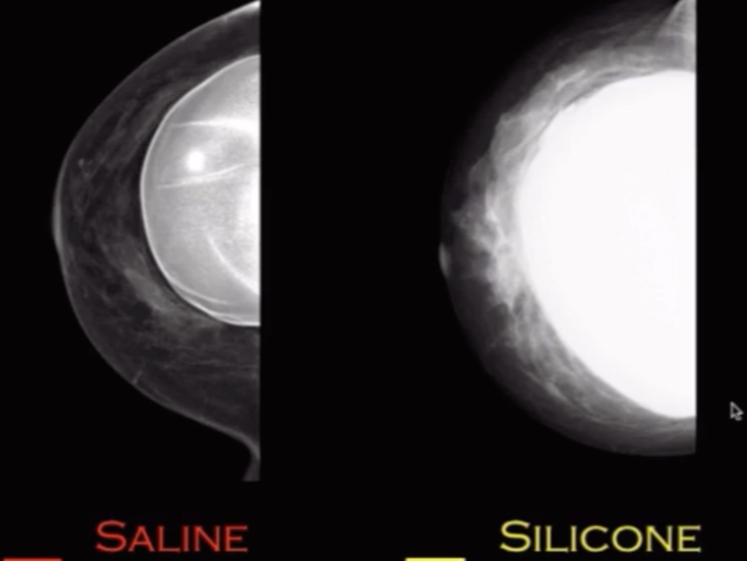
Saline Implant
Can see through them
Suspect rupture —> physical exam & mammogram
Will not form a capsule so no such thing as intracapsular rupture
Rupture doesn’t really matter from a management standpoint, cosmetic issue only
Silicone Implant
Cannot see through them
Suspect rupture —> start with US probably, will ultimately need MRI
Capsule
Body will form a capsule around the implant made of scar tissue and shit
Note that the actual container holding the silicone is not the capsule it is tissue itself that walls off around the implant that is the capsule
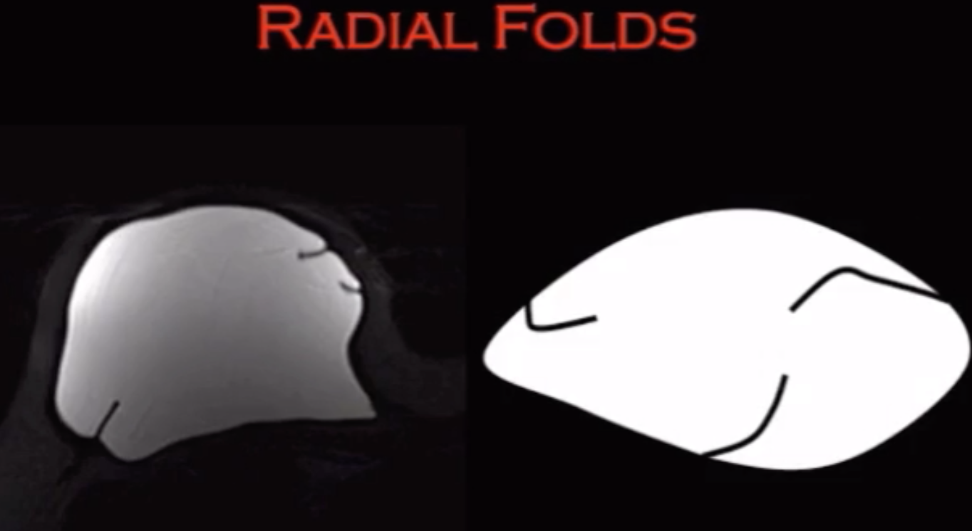
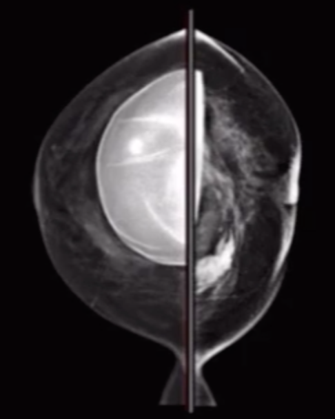
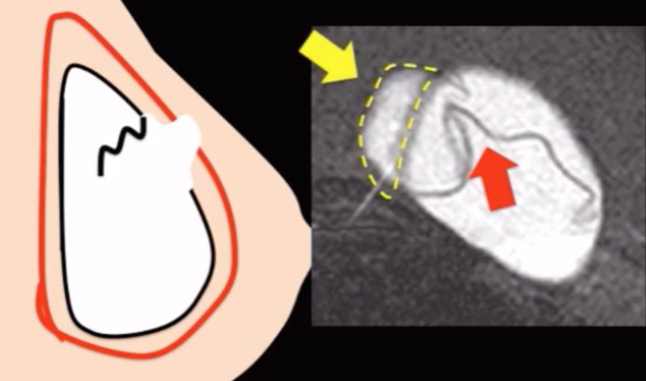
Intracapsular rupture (80%) - Image 1
Rupture of the container of the silicone implant but not the actual capsule (soft tissue capsule) itself
Called linguine appearance
Squiggle line will not have attachment to wall (radial fold will!)
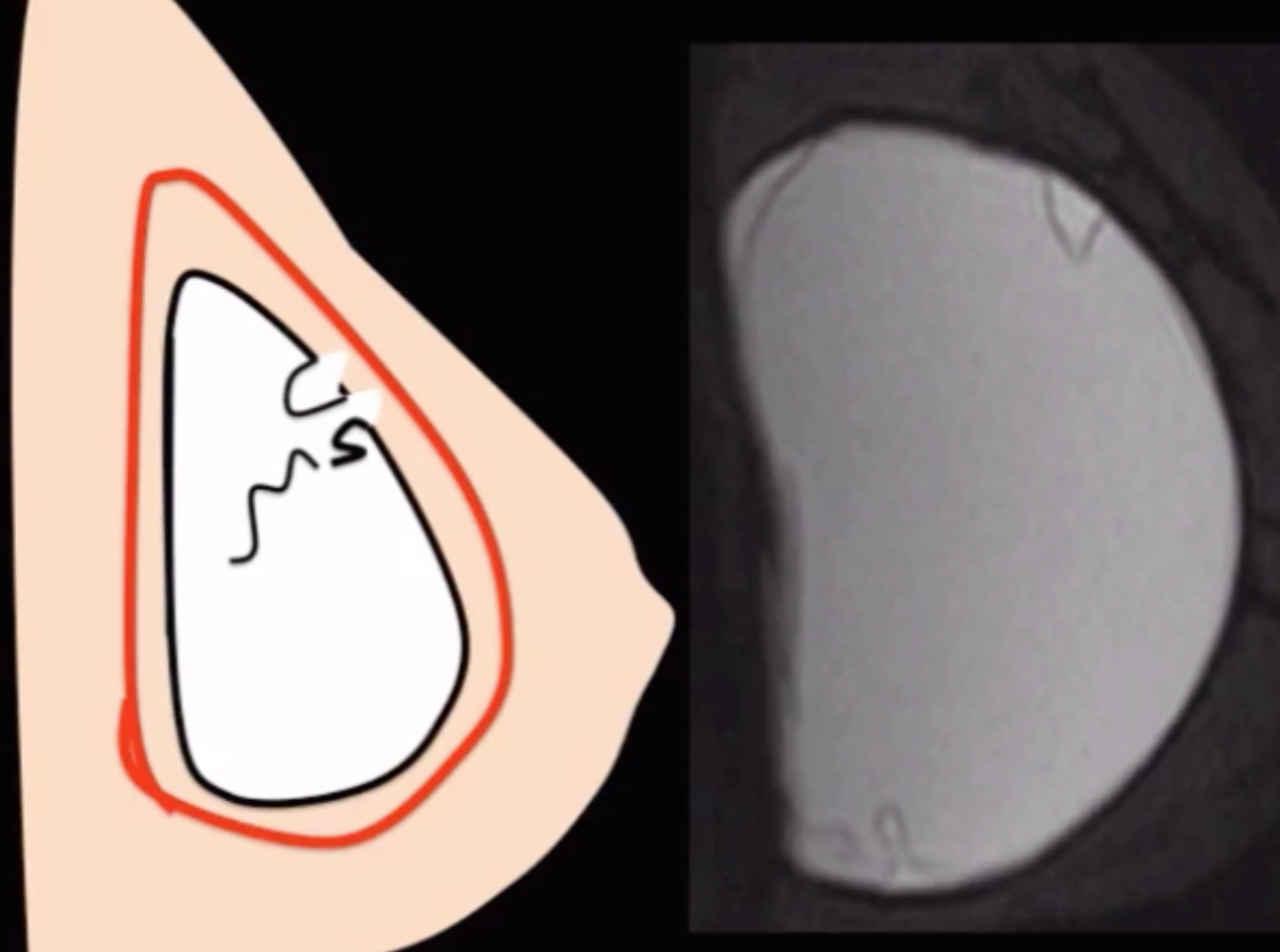
Extracapsular rupture (20%) - Image 2
Implies intracapsular + extracapsular
Cannot have extracapsular rupture alone
Injury violates the soft tissue capsule and container of the silicone implant
Snowstorm appearance on US - looks like dense blob
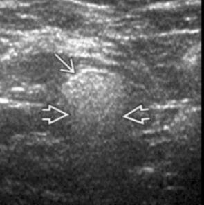
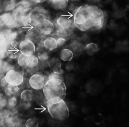
Silicone granuloma aka Silicone adenopathy aka Siliconeoma aka
See dense nodes on mammogram in pt with silicone implant
On US basically looks like snowstorm appearance, dense blob
Then need to recommend MRI to evaluate for implant rupture
Note: Gel bleed will present similarly
Normal transudation of micro-amounts of silicone through the container of the implant
Silicone Granuloma aka Silicone adenopathy aka Siliconeoma
See dense nodes on mammogram in pt with silicone implant
May have remote history of implant rupture and replacement/prior removal
On US basically looks like snowstorm appearance, dense blob
Hyperechoic nodules with posterior acoustic enhancement = silicone granuloma
Then need to recommend MRI to evaluate for implant rupture
Note: Gel bleed will present similarly
Normal transudation of micro-amounts of silicone through the container of the implant
Radial Fold
Basically the normal folds in the capsule of the breast implant
Mimics rupture
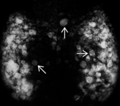
Silicone Injections
Looks like numerous, diffuse bilateral masses (almost like numerous oil cysts)
Will have snowstorm appearance too
Breast become hard and very dense
MR is really the only useful imaging modality in these women
Breast Reduction Surgery
Will see reorganization of breast tissue near the incision sites, bilaterally
Will present as thickened linear breast tissue near incision sites
Benign calcifications typically seen about 2 years after surgery
New calcs earlier than this should be investigated for malignancy
Typical incision locations
Inferior breast
Peri-areolar
Inframammary folds
References:

Trabecular Thickening
Thickening of the linear fibrous septae, inclduing Cooper ligaments
Typically caused by edema
Seems to appear similar to breast reduction changes but without hx of surgery obv