Basics / Search Pattern
TAVR Study
Read like normal CT chest with special additions
Items to specially evaluate
Aortic valve for calcifications
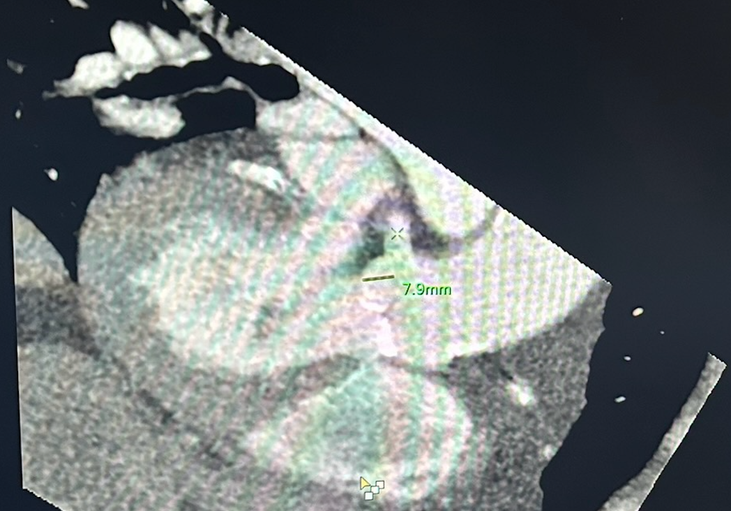
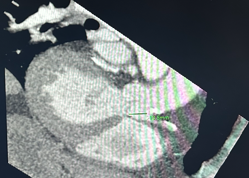
Looks at origin of coronary arteries and the distance of the RCA & Left main from the aortic valve so that when prosthetic valve is placed it does overlap on the coronary artery origin and occlude them
Femoral and iliac arteries to evaluate the arterial access site for the actual procedure
LV outflow tract angle
Aortic valve diameter
Steps
Read everything but the heart like normal
Read coronary calcium score like normal
Now the TAVR specific
Right click the first raw data picture (static looking image)
Click philips applications —> cardiac CT viewer —> click open when new tab opens
Will need to manipulate image so that the following screen shots can be obtained
MAKE SURE TO TAKE SCREENSHOTS OR YOU WILL HAVE TO REDO EVERYTHING
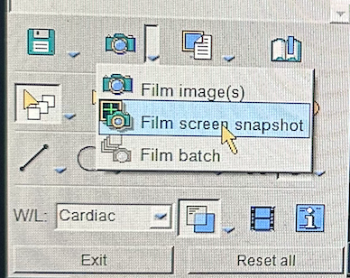
Bottom right corner —> camera drop down —> film screen snapshot
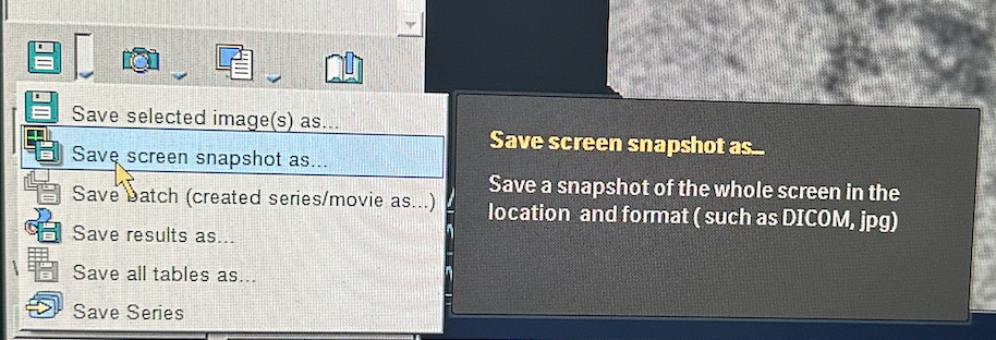
Then floppy disc save drop down —> save screen snapshot as —> new menu will open asking where to save —> PACS should be auto-selected but ensure that is what is selected and then click save
RCA + Aortic valve in same plane with measurement included (left image)
Left main + Aortic valve in same plane with measurement included (right image)
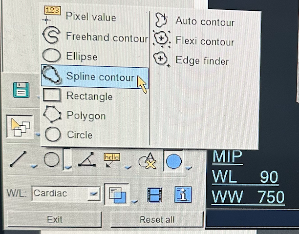
Circumference of the aortic valve can be measured
Use spline tool (bottom right corner —> circle tool dropdown menu —>spline contour
LV outflow tract with proximal aorta
Click the angle measurement in the bottom left and draw line from apex of LV to aortic valve, then second portion extending from valve along the plane of the ascending aorta
References: