Testicular Ultrasound
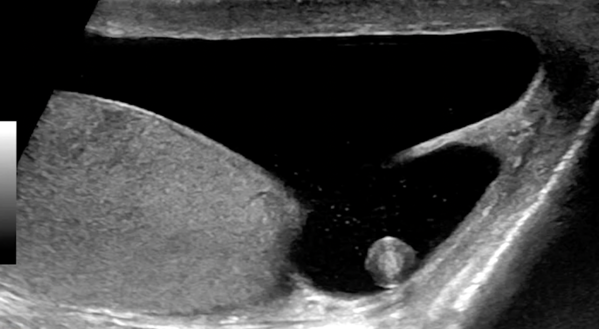
Testicular Appendage Torsion
Benign, managed conservatively
Look for round extra-testicular mass without doppler flow
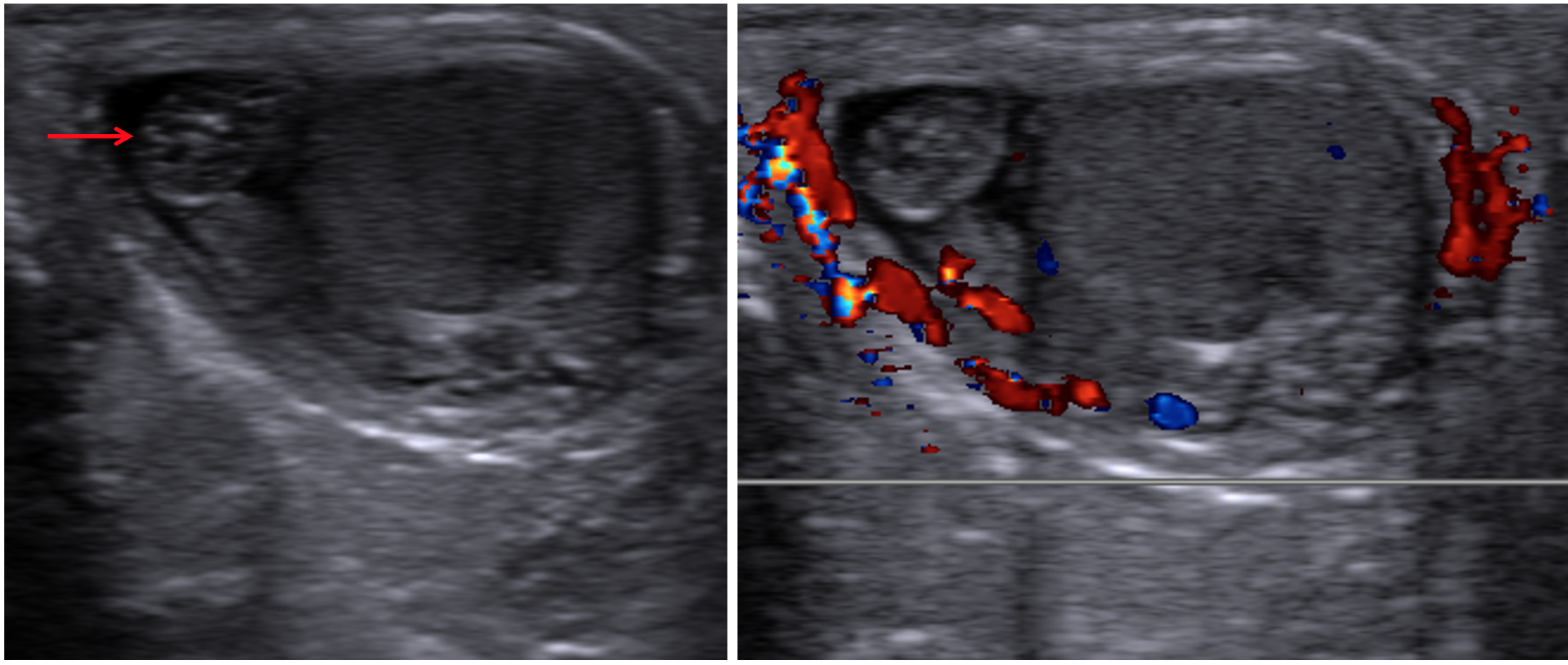
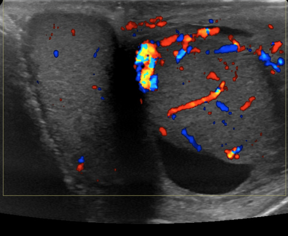
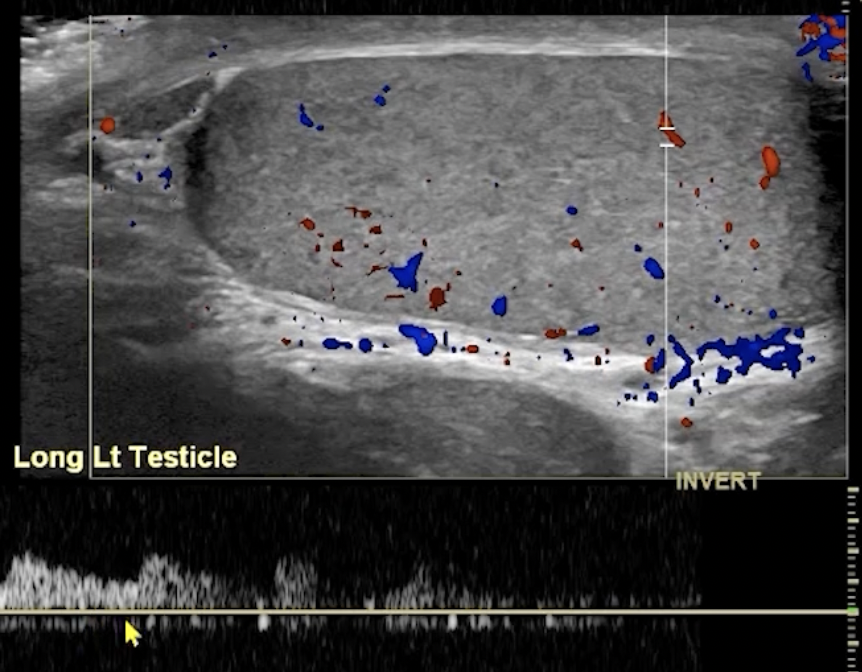
Epididymo-Orchitis
Inflammation of the epididymis and testicle
Presents as an enlarged and hyper-emic (increased blood flow) testicle/epididymis
May also have
Associated hydrocele
Check to make sure it is not septated —> if septations are present = pyocele
Hyper/hypo/heterogenous echotexture
Epididymal Cyst & Spermatocele
Both look like a simple cyst next to testicle
Anechoic with thin wall
Only can tell difference if you aspirate them and spermatocele will have sperm, the epididymal cyst will have simple fluid
Scrotal Pearl
Testicular appendage essentially twists off and just sits within the scrotum and the calcifies
Torsion
Look for lack of flow
Look for lack of attachment to scrotal wall, likely suspended in a hydrocele
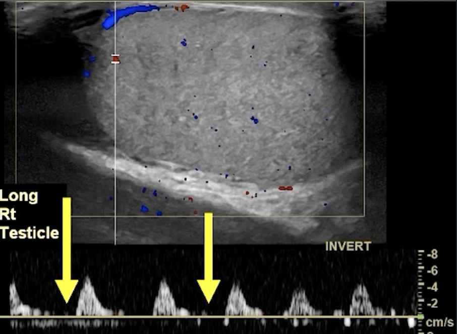
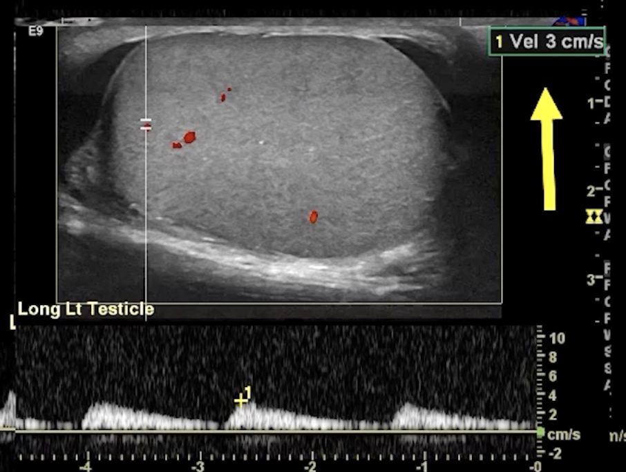
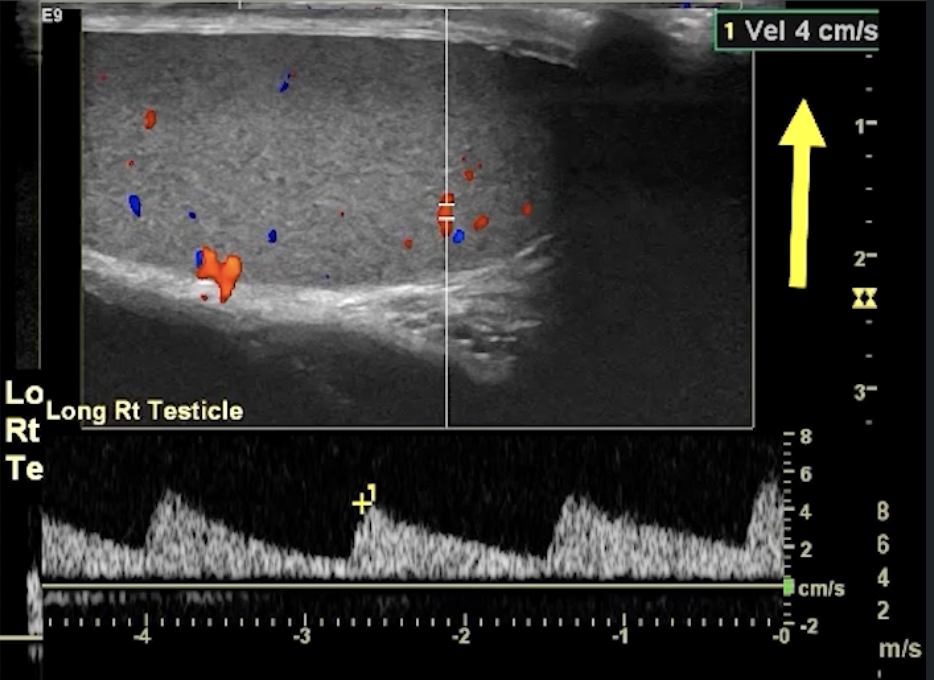
Partial Torsion
May have some blood flow
Decreased or reversal of flow in diastole (no line in between peaks) - indicates high resistance state (top images)
Blunted arterial peak (not as sharp as a point in systole) ( bottom images)
Abnormal
Testicular Infarction
Will appear as hypoechoic area in testis
Compare to other side to see what the normal echogenicity should be as if most of the testicle is infarcted then only the small area of normal testicle parenchyma may look bright and it would make it look like since the bright area is the minority that it is the abnormal part when that is not true
From direct injury to vasculature or thrombus
Do not need bell clapper to have this
Small testicle
Unilateral
Trauma
Infarction (i.e. torsion, hernia repair, infectious epididymo-orchitis, trauma)
Typically small, heterogenously hypoechoic, may have calcs
Bilateral
Hypogonadism
Zinner Syndrome
Triad of Wolffian duct anomalies
Triad of
Unilateral renal agenesis
Ejaculatory duct obstruction
Seminal vesicle cyst
Male urethra cancer
Anterior urethra mets to the inguinal nodes
Posterior urethra mets to the iliac and obturator (pelvic) nodes
Normal
Undescended Testicle
Usually will see in younger patients
Can present as a big pelvic mass with associated neoplasm if chronically undescended and unknown
Increased risk of malignancy in both testicles not just the descended one
Increased risk of infertility
Cancer
Dilated Rete Testes
Normal variant of dilated tubules
Associated with epididymal cysts
Post-Vasectomy changes
Congested appearance of the seminal vesicles
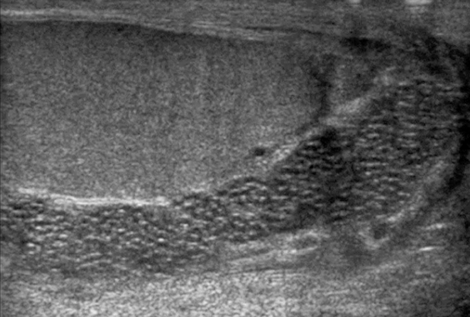
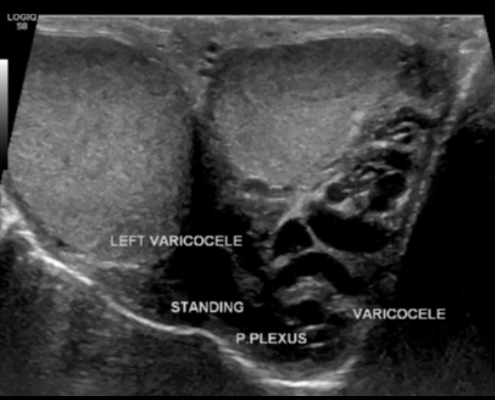
Varicocele
Dilated vessels outside of testicle
Should have some doppler flow
If suspect this, have patient valsalva while ultrasounding and should see increased flow within the vessels
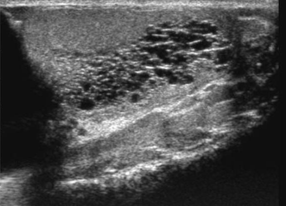
Testicular Microlithiasis
Largely a benign finding seen in kids
Can follow with US
Used to be thought there was association with malignancy but not anymore
References: