Spine
Os Odontoideum
Anatomic variant associated with atlanto-axial instability
Associated with Marquio syndrome (Mucopolysaccharidosis type 4)
Klippel-Feil Syndrome
Poor segmentation of embryo in weeks 3-8 results in multiple deformities, most notable in cervical spine
Triad
Short neck (secondary to cervical fusion)
Poor neck mobility (secondary to cervical fusion)
Low hairline
Multilevel cervical fusion is the major finding
Associated spine issues such as disc herniations, scoliosis, etc. are also seen, likely as sequelae of having bad cervical spine
Other important irregularities associated with K-F
Aortic arch and great vessel anomalies
GU tract anomalies (unilateral renal agenesis)
Sprengel deformity
Unilateral elevation of scapula
Classically has omovertebral bone - osseous/cartilagenous/fibrous connection of scapula to spine
Cavendish & Rigault classifications further characterize but too much detail to care
If you see this deformity you should look for other anomalies associated with K-F (cardiac, GU, etc) !
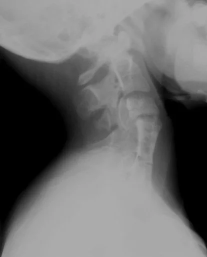
Left:
Multilevel cervical fusion shown well
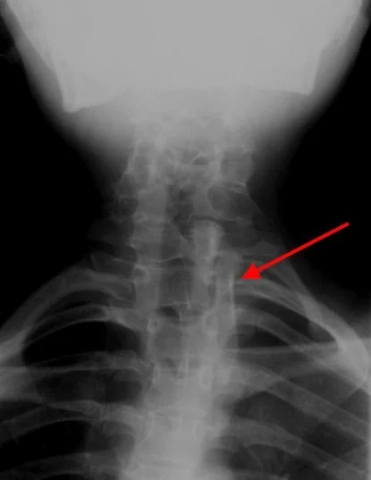
Right:
Demonstrates left sided omnovertebral bone adjacent to C7-T3
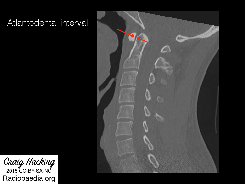
Atlantodental interval
Basically space between anterior aspect of dens and posterior aspect of C1
Normal
< 3 mm on XR
< 2 mm on CT
Widening of the atlantodental interval is highly suggestive of transverse ligament injury
Sources:
Case courtesy of Vincent Tatco, Radiopaedia.org, rID: 40862 (K-F case)
Case courtesy of Craig Hacking, Radiopaedia.org, rID: 40491 (atlantodental case)