Skull Anatomy
Skull Foramen
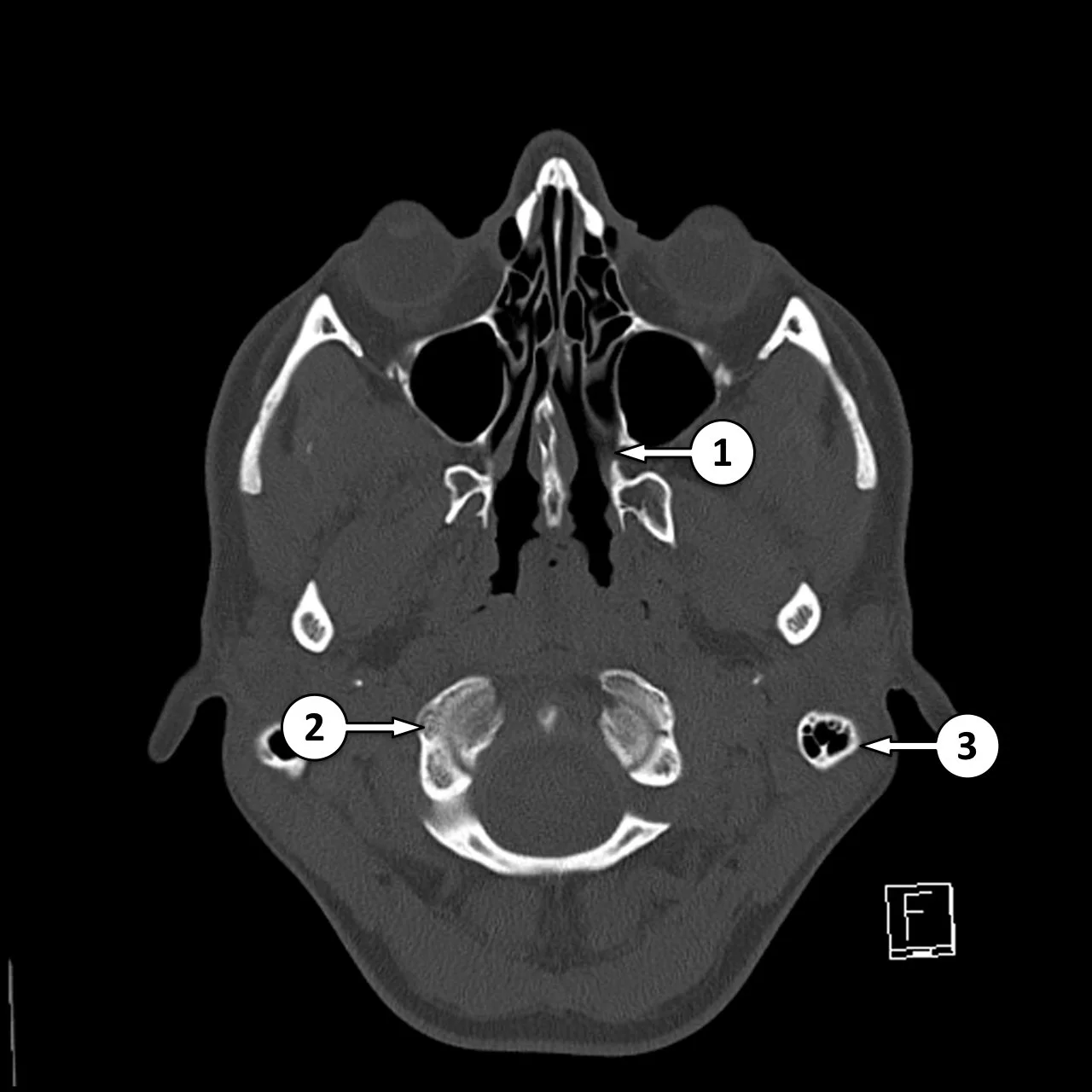
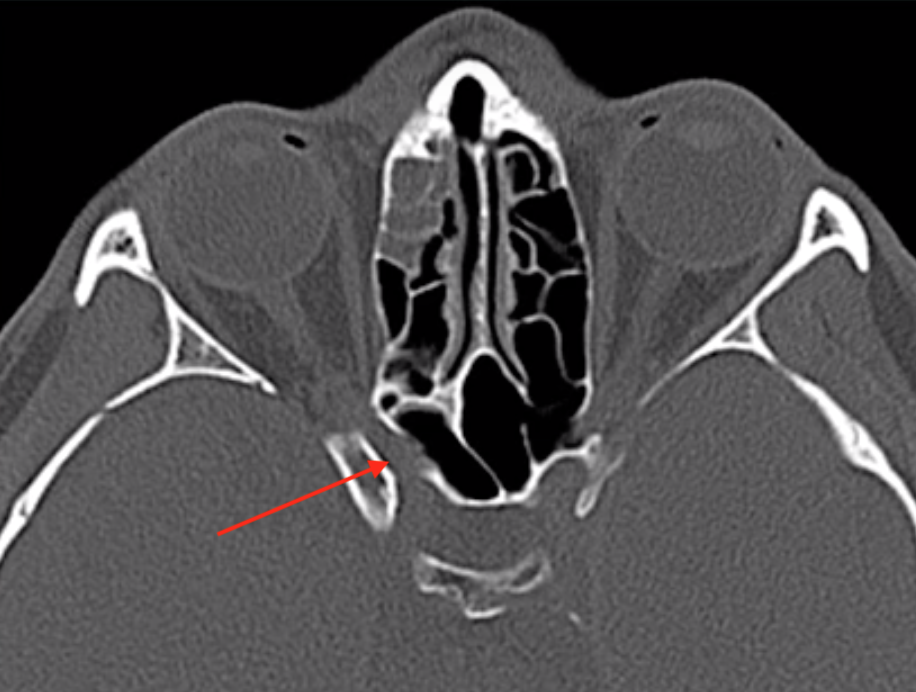
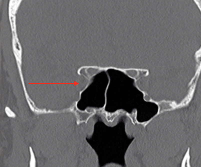
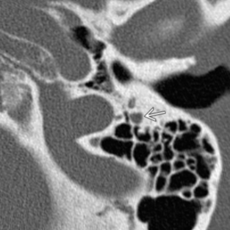
Sphenopalatine Foramen
#1 in image to the right
Allows passage of nasal cavity and pterygopalatine fossa
Transmits
Sphenopalatine artery & vein
Nasopalatine nerve
Posterior superior nasal nerves
Clinical relevance
Because nerves travel through here, it allows a possible passageway for peri-neural spread of malignancy from the nasal cavity to deeper structures
Juvenile Angiofibromas
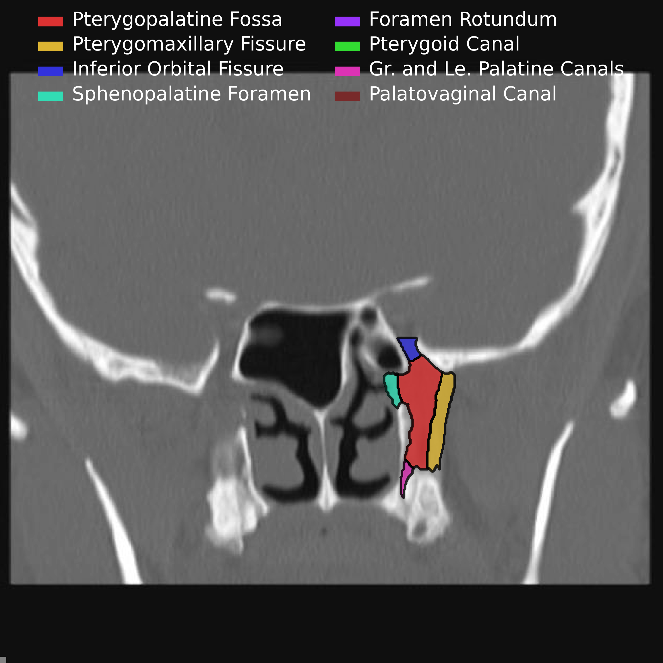

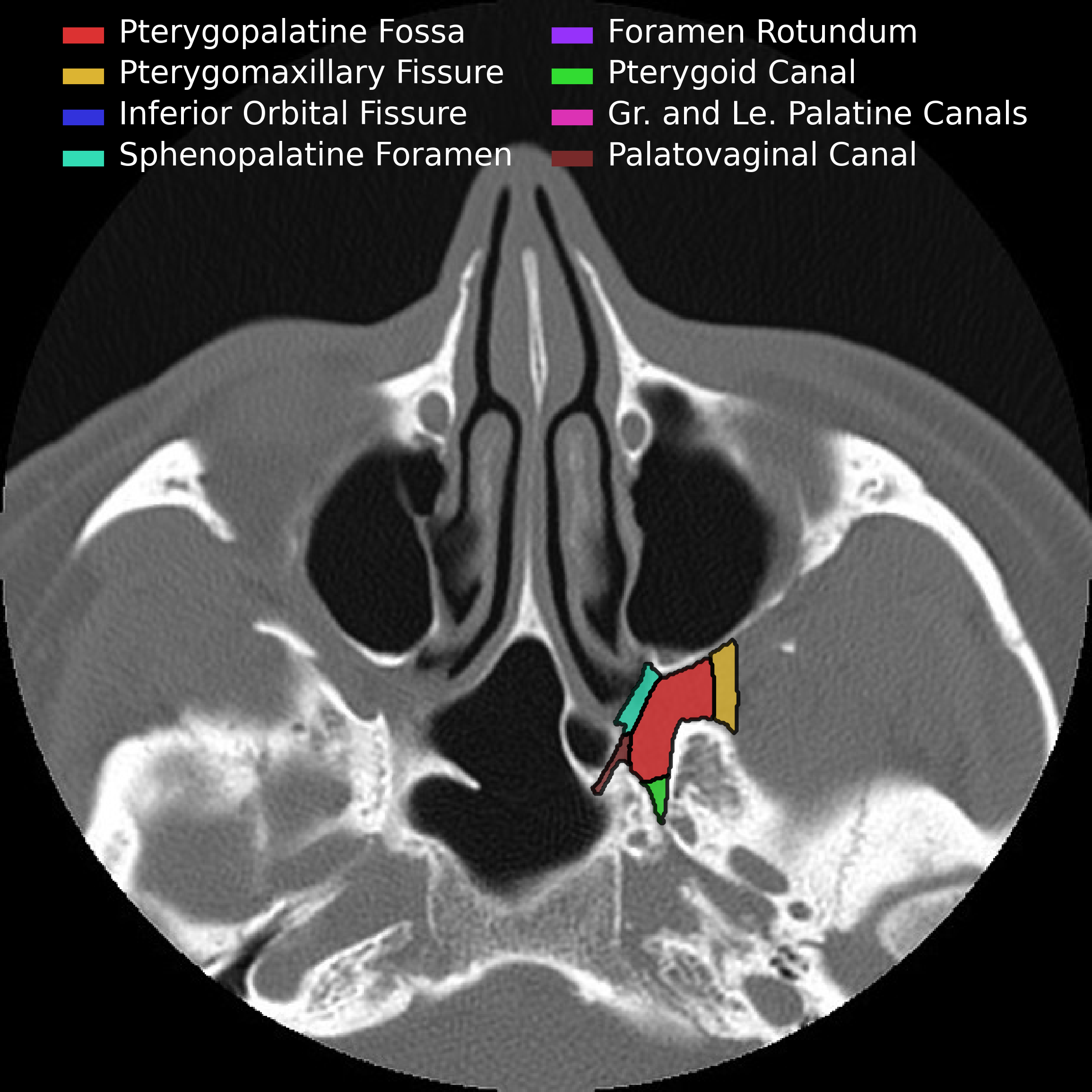
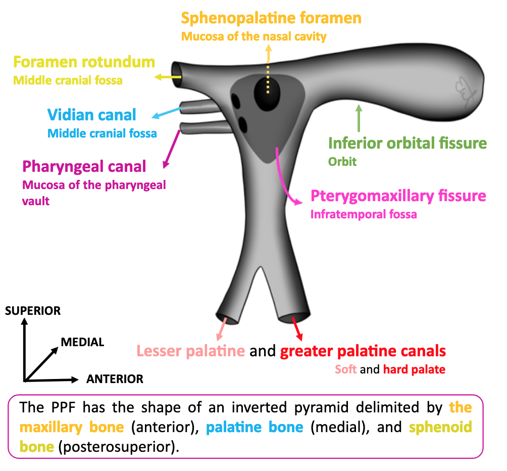
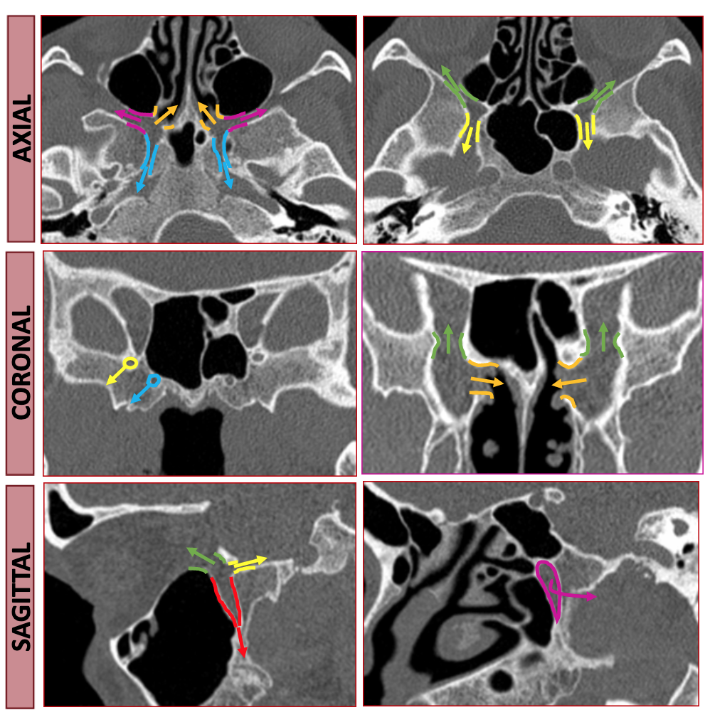
Pterygopalatine Fossa
Should think of pterygopalatine fossa as one of the major intersections of the face/skull
Contents:
Pterygopalatine ganglion
Terminal aspect of Maxillary artery (& descending palatine artery branch)
Emissary veins
Maxillary division of CN V, via foramen rotundum
Pterygoid canal nerve
The following pictures are a fantastic way to visualize the borders rather than just memorizing them which is a sure fire way to forget them by tomorrow.
Anterior & Superior - communicates with inferior orbital fissure
Inferior - Greater & lesser palatine canals
Communicates with palate
Lateral - Pterygomaxillary fissure
Communicates with masticator space
Medial communication - Sphenopalatine foramen & Palatine bone
Communicates with nasal cavity
Transmits
Sphenopalatine artery
Posterior superior nasal nerves & nasopalatine nerve
Posterior
Posterior-medial - Palatovaginal canal
Communicates with nasopharynx
Transmits pharyngeal nerve and pharyngeal branch of maxillary artery
Posterior-superior - Foramen rotundum
communicates with Meckel cave & Cavernous sinus
Posterior-inferior - pterygoid canal (aka vidian canal)
Communicates with middle cranial fossa
Transmits vidian nerve, artery and vein
Pterygopalatine Fossa
Important space because allows spread of malignany

Skull Foramen
High yield and easy points if you know them
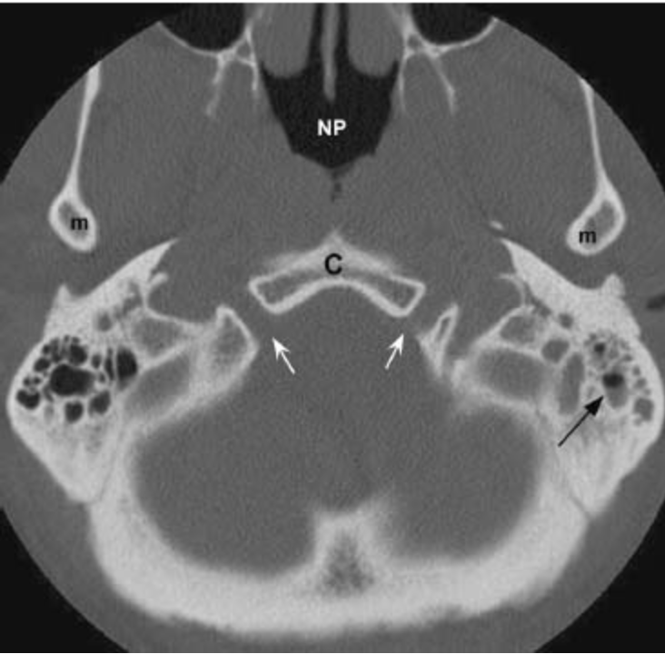
Foramen Lacerum
Does not transmit any structures
Is the cartilaginous floor of the anteromedial horizontal carotid canal in the temporal bone.
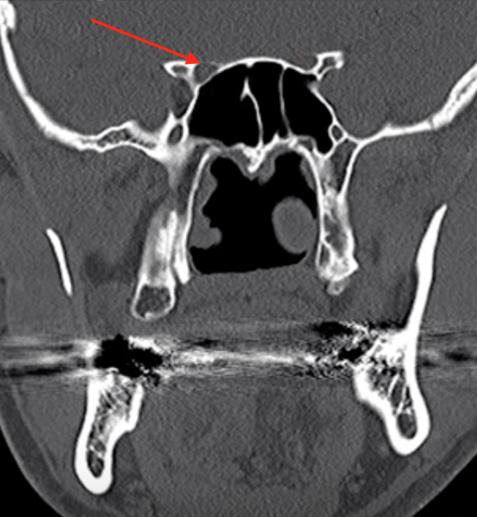
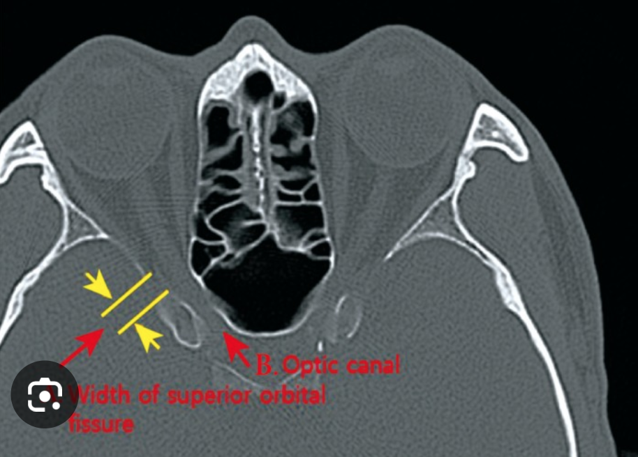
Optic Canal
Structures passing through here:
CN 1
Ophthalmic artery
Superior Orbital Fissure
Structures passing through here:
CN 3, 4, 5-V1, 6
Superior ophthalmic vein
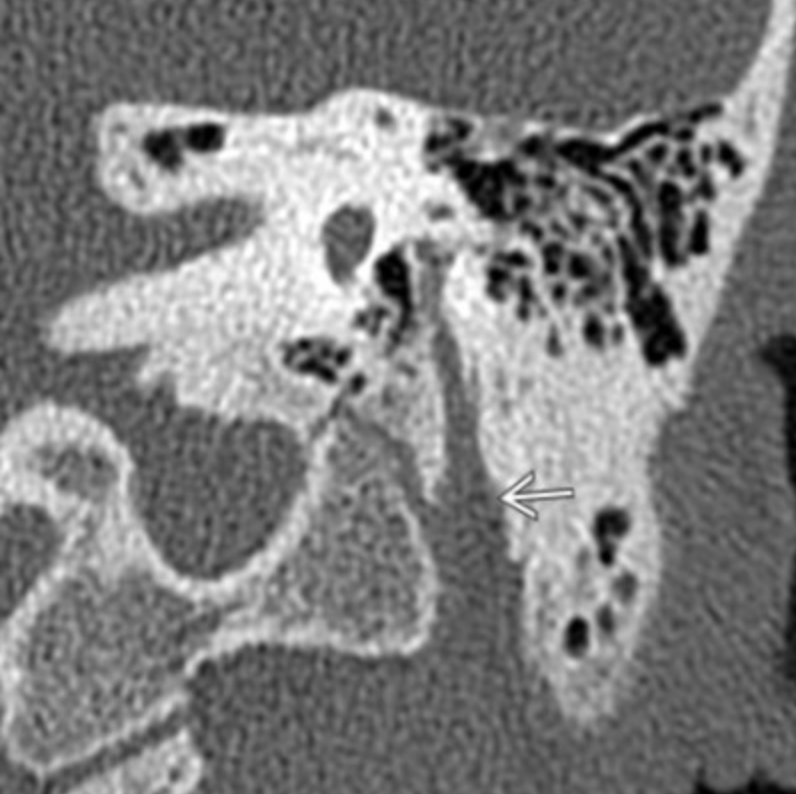
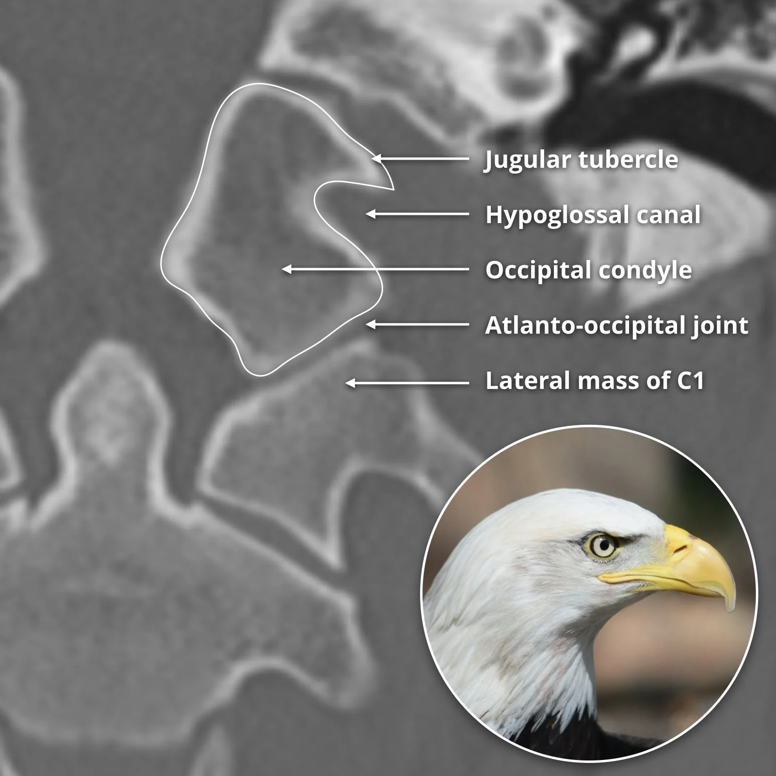
Hypoglossal Canal
Hypoglossal Nerve (CN12)
Motor to tongue
Note palatoglossal nerve is only extrinsic tongue muscle not innervated by 12 (it is innervated by CN 10)
Stylomastoid foramen
Facial nerve exits the base of the temporal bone through tstylomastoid foramen
Only motor portion of CN7 exits the stylomastoid foramen
CN 5 - Trigeminal Nerve
Trigeminal ganglion lies in Meckels cave which is a depression in the temporal bone just lateral to the cavernous sinus
Branches
Ophthalmic - V1
Exits via superior orbital fissure
Terminal branches
Frontal nerve
Lacrimal nerve
Nasociliary nerve
Controls blinking reflex when shit comes toward you (corneal reflex)
Maxillary - V2
Exits via foramen rotundum
Multiple terminal branches including the inferior orbital nerve
Mandibular - V3
Exits via foramen ovale
Terminal branches
Buccal nerve
Inferior alveolar nerve
Auriculotemporal nerve
Lingual nerve
General sensory to anterior 2/3 of tongue (not taste)
Peri-neural Spread
Inferior orbital nerve (branch of maxillary V2)
CN 7 - Facial Nerve
Special Features
Taste to anterior 2/3 of tongue
Noise dampening
Lacrimation
Branches
Chorda tympani
Most inferior intra-temporal branch
Therefore first to be affected by parotid gland lesion extendning intracranially
Stapedius branch
Dampens noise
Just proximal to the chorda tympani
Greater superficial petrosal nerve
To lacrimal gland
Proximal to the stapedius branch
Exits skull at stylomastoid foramen where it enters the parotid space
Nerve will pass lateral to the retromandibular vein
Temporal bone segments
IAC segment: Porus acusticus to IAC fundus; anterosuperior position above crista falciformis
Labyrinthine segment: Connects fundal CNVII to geniculate ganglion (anterior genu)
Tympanic segment: Connects anterior to posterior genu, passing under lateral semicircular canal
Mastoid segment: Inferiorly directed from posterior genu to stylomastoid foramen
Cranial Nerves
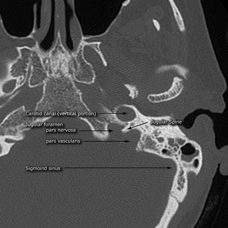
Jugular Foramen
Divided into two separate pars via the jugular spine
Pars Nervosa
Anterio-medial
Transmits CN 9
Pars Vascularis
Posterio-lateral
Transmits CN 10 + CN 11 + Jugular vein
References:
Case courtesy of Craig Hacking, Radiopaedia.org, rID: 62425 (CT images)
Case courtesy of Reuben Schmidt, Radiopaedia.org, rID: 177576 (CT with colored sphenopalatine fossa)
https://e-acfs.org/m/journal/view.php?number=555 (statistical analysis of superior orbital fissure in korean adults using CT)
Jugular foramen picture - Case courtesy of Frank Gaillard, Radiopaedia.org, rID: 35926
Hypoglossal canal - Case courtesy of Frank Gaillard, Radiopaedia.org, rID: 57699