Prostate MR
General
African American most common population
Most common primary malignancy in men, second most common cause of death
IV glucagon commonly given to slow bowel peristalsis so there is less motion artifact
Contrast typically not needed
The MRI is typically for staging although increasingly used to screen
PSA
Normal = 1-4
>4 is abnormal
Prostate Cancer
95% = adenocarcinoma
70% = Peripheral zone
Majority will be area of hypointensity
Mucinous adenocarcinoma (5%) will be area of increased signal in peripheral zone
30% = Central and anterior zone
Note if there was a recent biopsy then will have area of high T1 signal because of blood
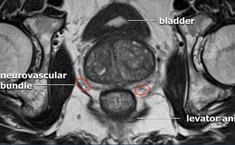
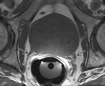
Neurovascular bundle
The straight lines that look like they are extending laterally from the posterior border of the prostate
If they look hazy then consider involvement, doesn’t look like definitive invasion can just have haziness around it
Black arrows in image below
T Stage
T1: Clinically localized [tumor not palpable on digital rectal exam (DRE)]
T2: Tumor confined to prostate
T3: Locally invasive beyond prostatic capsule
T4: Tumor is fixed or invades adjacent structures other than seminal vesicles, such as external sphincter, rectum, bladder, levator muscles, &/or pelvic wall
References: