Pediatric Chest Radiology
Scimitar Syndrome
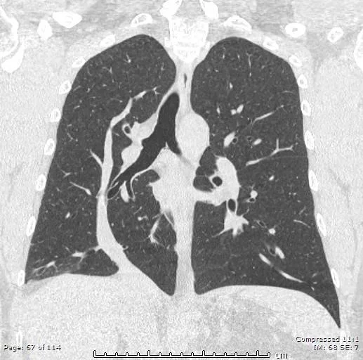
Congenital malformation where a hypoplastic lung is drained by its own pulmonary vein into systemic venous circulation (most commonly the IVC, less commonly RA or other sites)
Results in an acyanotic left to right shunt
Normal pulmonary veins get blood from the lungs and bring back to the left atrium
Here we have a rogue pulmonary vein that is bringing blood from an isolated right lung segment back to the IVC.
The left heart gets oxygenated blood. The right heart has deoxygenated blood. Here (i believe) the blood from teh rogue lung goes back to the systemic venous system (to the IVC) and is already oxygenated since it came from the rogue piece of lung which is why we therefore have an acyanotic left to right shunt.
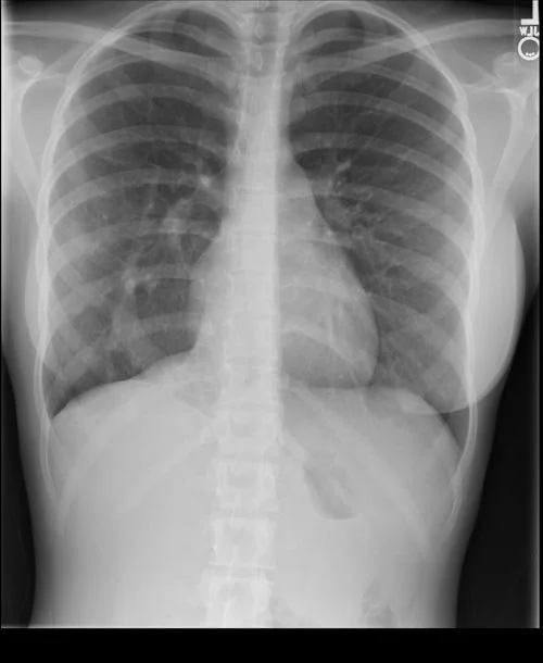
Scimitar is turkish word for a sword which is what the anomalous draining vein looks like on imaging - those swords the bad guys from Aladdin have
Included in the spectrum of PAPVR
Really only ever seen on the right side
Imaging -
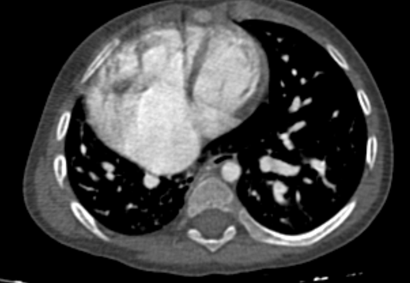
On CT looks like a tension pneumothorax without the collapsed lung
On XR look for the scimitar - aka thick vessel looking structure on the right side
Associated with multiple things -
Horseshoe lung
Cardiac anomalies
Pulmonary sequestration
Multiple others (vertebral, GU tract anomalies)
References:
Case courtesy of The Radswiki, Radiopaedia.org, rID: 11917 (Scimitar)
Case courtesy of Husam Hussein Yaseen, Radiopaedia.org, rID: 75133