Pancreas
Acute Pancreatitis
Pancreas is one of the fattier organs in the body
Typically the pancreas appears hyper-echoic (bright) on ultrasound because it contains a lot of fat. (remember in hepatic steatosis the liver also appears bright on US). So in acute pancreatitis the pancreas will appear hypo/iso-echoic (dark) as there will be edema within and around the pancreas which makes it dark as fluid appears dark on US.
Pancreas US
Typically bad to diagnose anything
Pancreatic duct dilatation
Pancreatic duct > 3mm
Needs follow up CT or MRCP
Double duct sign
Abnormally dilated pancreatic and biliary duct = no bueno = concern for mass —> get CT
Pncreatitis
US typically just to look for cause, specially a stone
Pancreas will have decreased echogenicity than normal due to the edema - pancreas normally very echogenic
Pay close attention to portal and splenic vein for thrombosis
Pancreatic necrosis —> pancreas will appear hyperechoic
Pancreatic Neuroendocrine Tumors
Masses that arise from pancreatic endocrine cells (islets of langerhans cells)
Mets to liver if they metastasize
Types
Insulinoma (most common)
Gastrinoma
Non-functioning
Solid and Pseudopapillary Neoplasma (SPEN)
Well defined
Typically in tail of pancreas
Hypoechoic on US
Solid mass with hemorrhagic and necrotic focus
Low risk but still some risk of malignancy
Women, <35
Pancreatic Masses
Serous Cystadenoma
Innumerable microcysts in a mass (Described as honeycomb)
>60 years old typically
Prefer head of pancreas
No ductal dilatation
Benign, no need for treatment

Pancreatic Neuroendocrine Tumor
Look for hypoglycemia
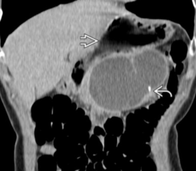
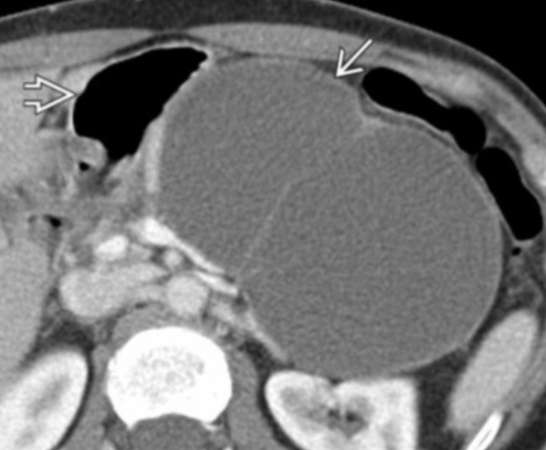
Mucinous Cystadenoma
Less than 6, large (2cm+) cystic lesions in a mass
Middle aged women
Prefer tail of pancreas
No ductal dilatation
Peripheral calcifications possible
Cystic Fibrosis
Typically atrophic fatty pancreas
Can also have numerous pancreatic cysts sometimes with calcs
But in a young person they should not have chronic pancreatitis already
Biliary manifestations
Micro-gallbladder, I mean really fucking small
PSC appearance of the biliary ducts
Distal intetsinal obstruction
Absence of seminal vesicles in males
Other
References: