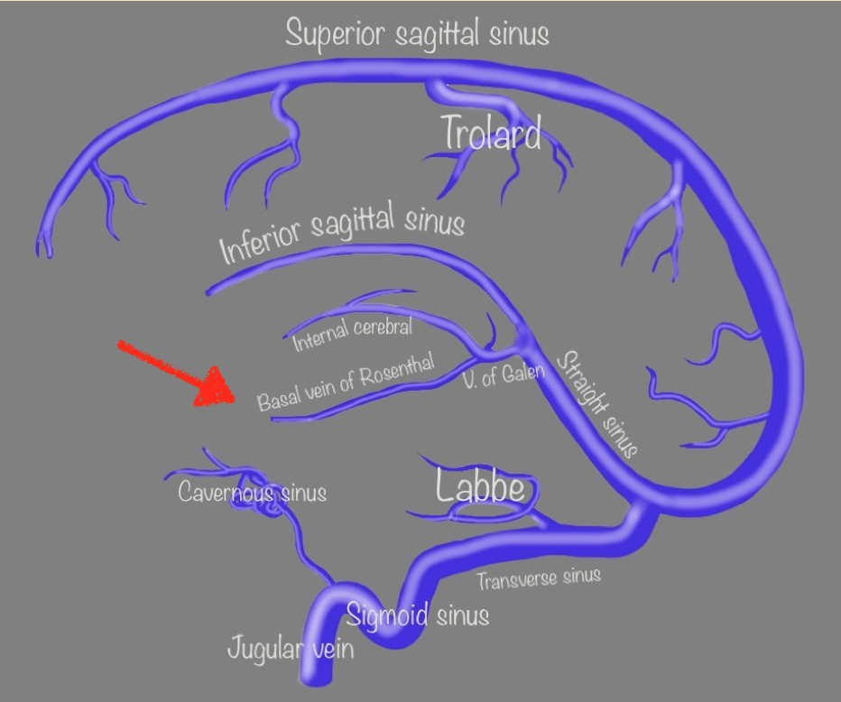
Neurovascular Venous Anatomy
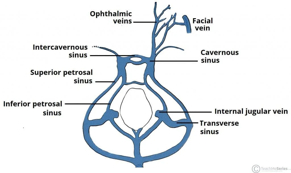
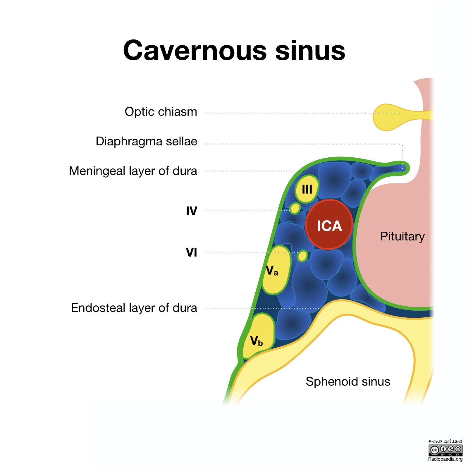
Cavernous Sinus
Contents (O TOM CAT) (Top to Bottom)
O - Oculomotor nerve (CN 3)
T: Trochlear nerve
O: Ophthalmic branch of CN 5
M: Maxillary branch of CN 5
C: Internal carotid artery
A: Abducens nerve
T: Trochlear nerve (again)
Pathology
Cavernous sinus thrombosis
Carotico-cavernous fistula
Mass of cavernous sinus (many possibilities)
Schwannoma
Meningioma
Pituitary macroadenoma
Hemangioma
Local mass spread (nasopharyngeal, perineural spread from other area)
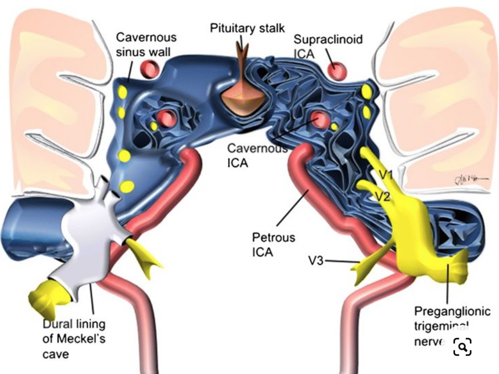
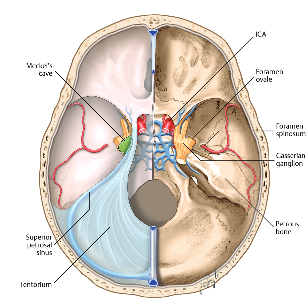
Meckel’s Cave
Sits posterior and lateral to cavernous sinus
ICA (lacerum & petrous segments) run underneath the cave
Pathology
Broadly divided into intrinsic and extrinsic lesions
Extrinsic lesions
Destroy surrounding bone trying to get into cave
Metastasis
Chordoma
Chondrosarcoma
Intrinsic lesions
Expand Meckel’s cave trying to get out of cave
Schwannoma (most common cause of cave mass)
Meningioma
Idiopathic intracranial HTN
References:
Case courtesy of Frank Gaillard, Radiopaedia.org, rID: 54907
Tushar Jha, H Jeffrey kim, and Walter C Jean (Skull Base Surgery: Strategies)