Neuro Vascular Anatomy
Circle of Willis
Rare to have complete circle of willis
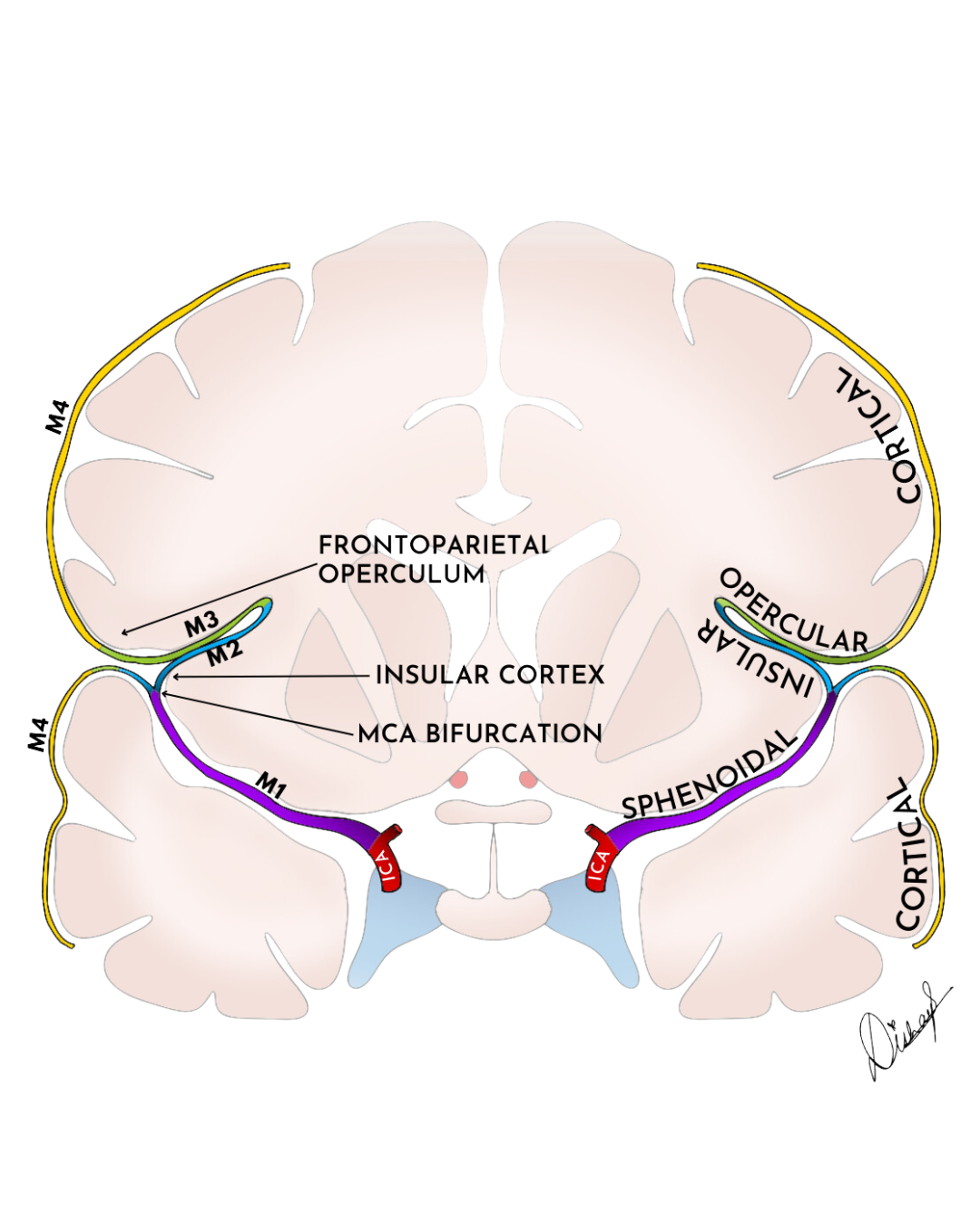
Middle Cerebral Artery (MCA)
Hyperdense MCA sign
Hyperattenuation of MCA on non-con CT
Earliest sign of ischemia in MCA stroke
Indicates presence of thromboembolism
M1: end of ICA to genu adjacent to limen insulae
Lenticulostriate are branches here
M2: limen insular to circular sulcus of insula
Divides into superior and inferior trunks
Superior M2 territory includes Broca’s areas
Inferior M2 segment includes Wernickie’s area
M3: circular sulcus of insula to superior surface of sylvian fissure
M4: superior surface of sylvian fissure to terminal end in cortical territory
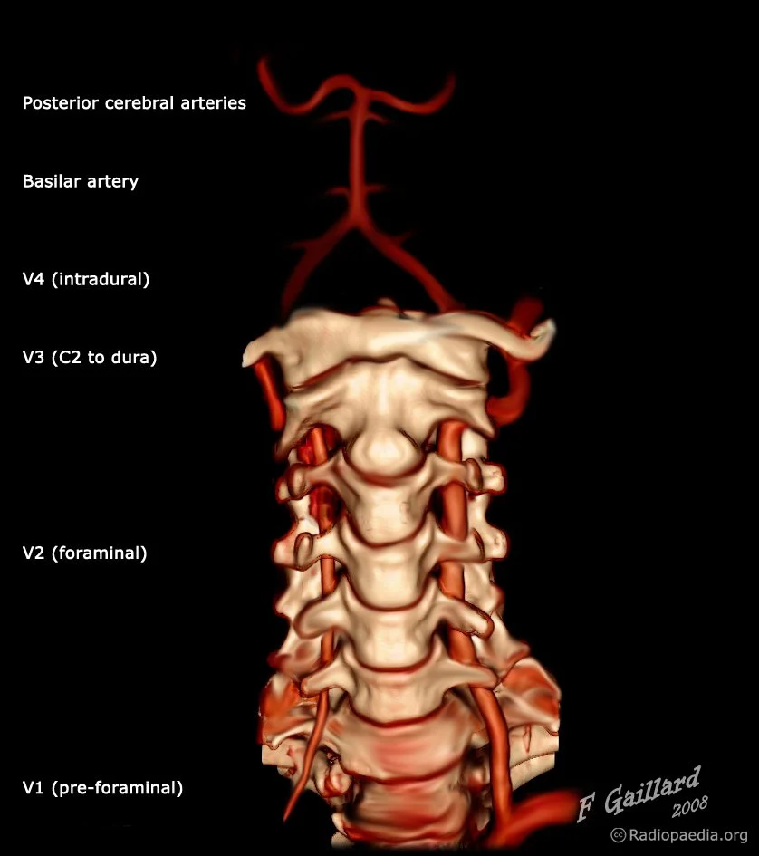
Vertebral Arteries
V1 = origin off subclavian to the vertebral foramen
V2 = intra-foraminal portion to the level of C2
V3 = C2 to dura
V4 = Intradural portion to the formation of basilar artery
PICA
Origin is highly variable
Most commonly off of vert just before basilar artery
PICA aneurysm rupture more commonly results in intraventricular hemorrhage + hydrocephalus > SAH alone
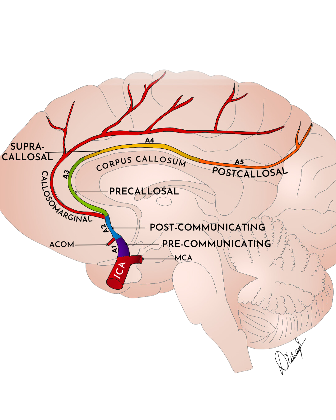
Anterior Cerebral Artery (ACA)
A1 = origin off ICA to Anterior communicating artery
A2 = Acomm to genu/rostrum intersection
A3 = Originates after callosomarginal artery branches to posterior turn around genu of corpus callosum
A4 = Superior anterior aspect of corpus callosum to coronal suture
A5 = Coronal suture and posterior until terminal end
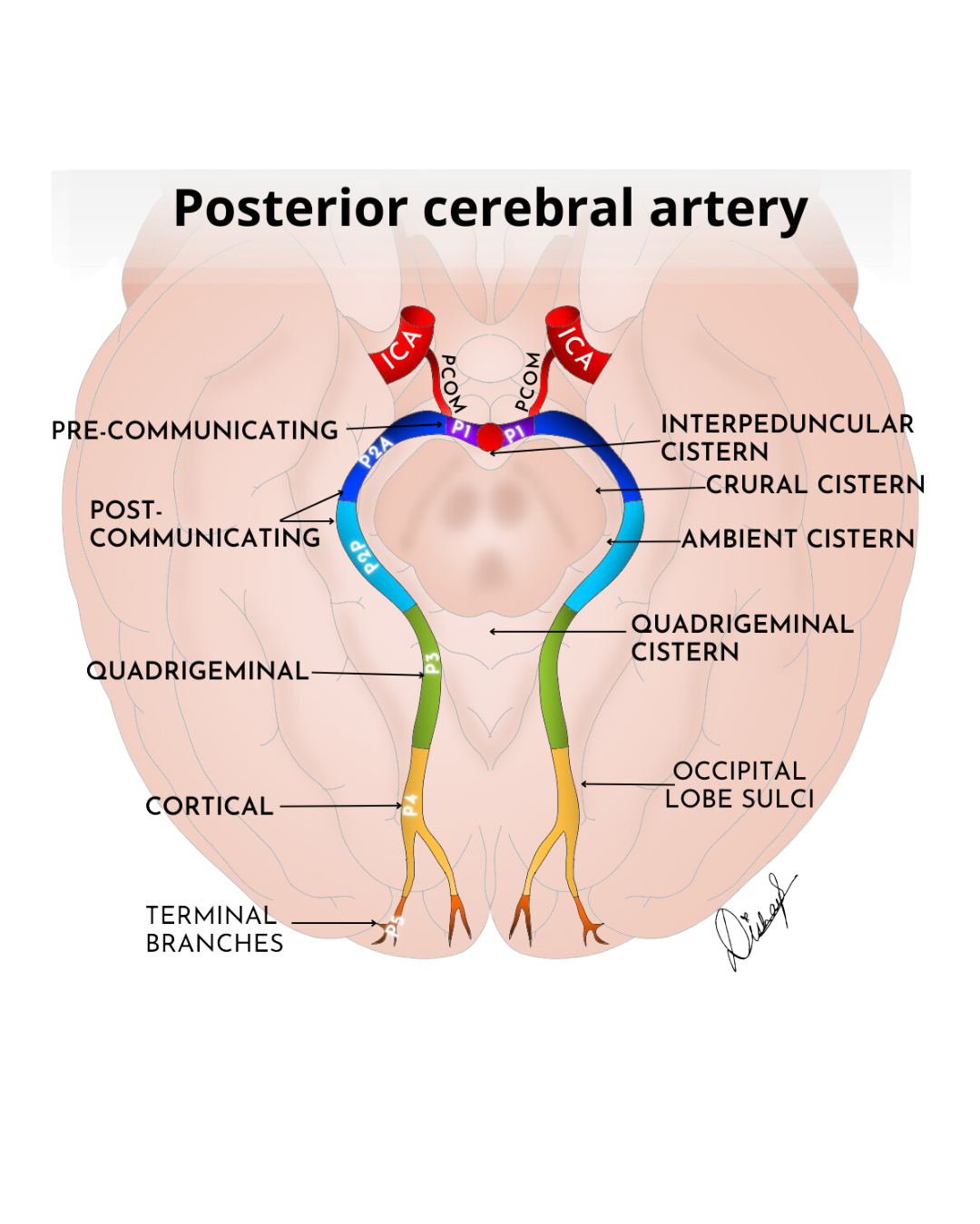
Posterior cerebral artery (PCA)
P1 = Basilar artery to PComm
P2 = Crural cistern to quadrigeminal cistern
P3 = Quadrigeminal cistern to occipital lobe
P4 = Occipital lobe sulci to terminal branches
P5 = Terminal branches
Fetal Origin of PCA
When PComm is bigger than P1 of PCA and supplies majority of blood to the P2 & distal segments of PCA
PICA - Posterior Inferior Cerebellar Artery
Arises from V4 segment of the vertebral artery just proximal to formation of the basilar
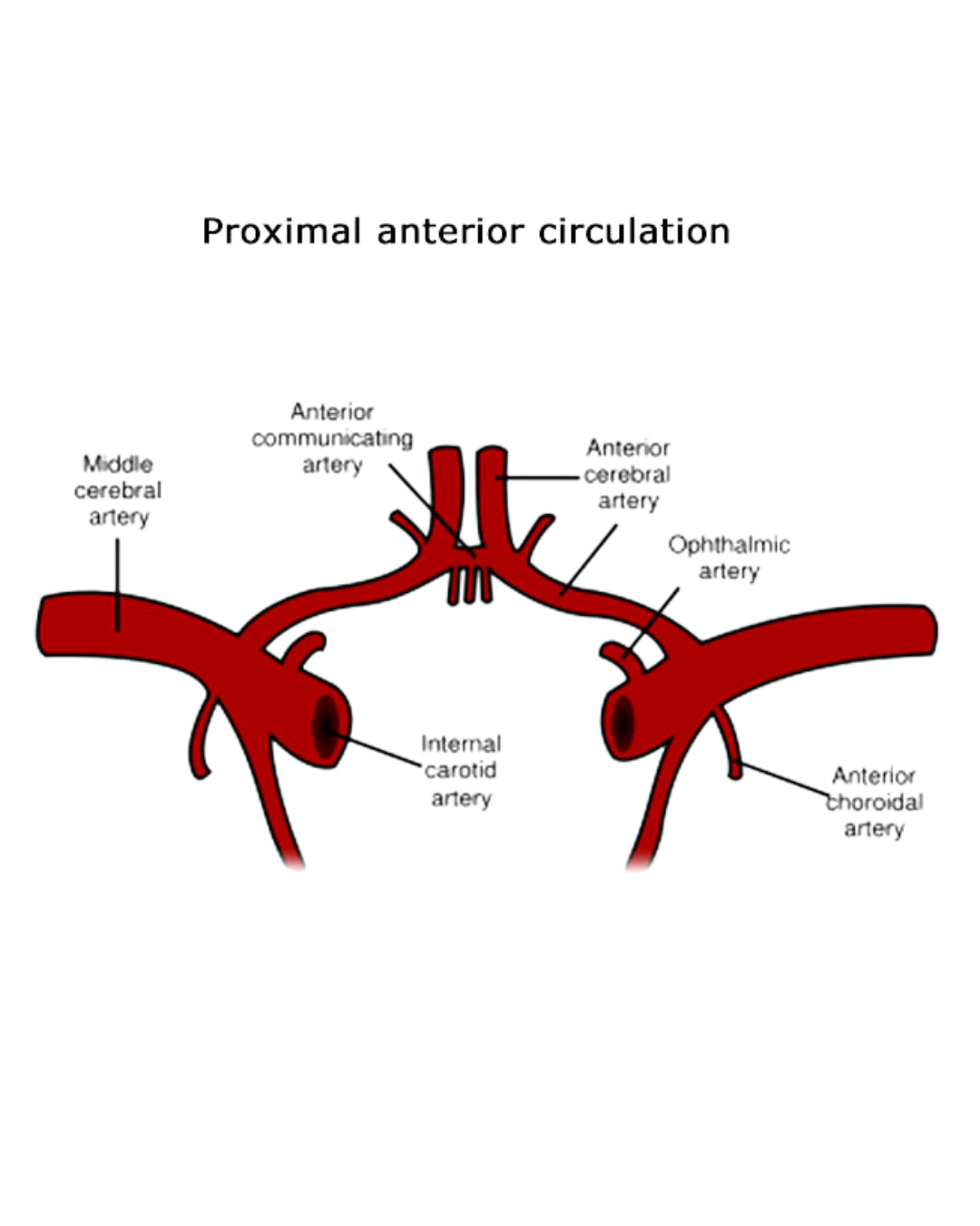
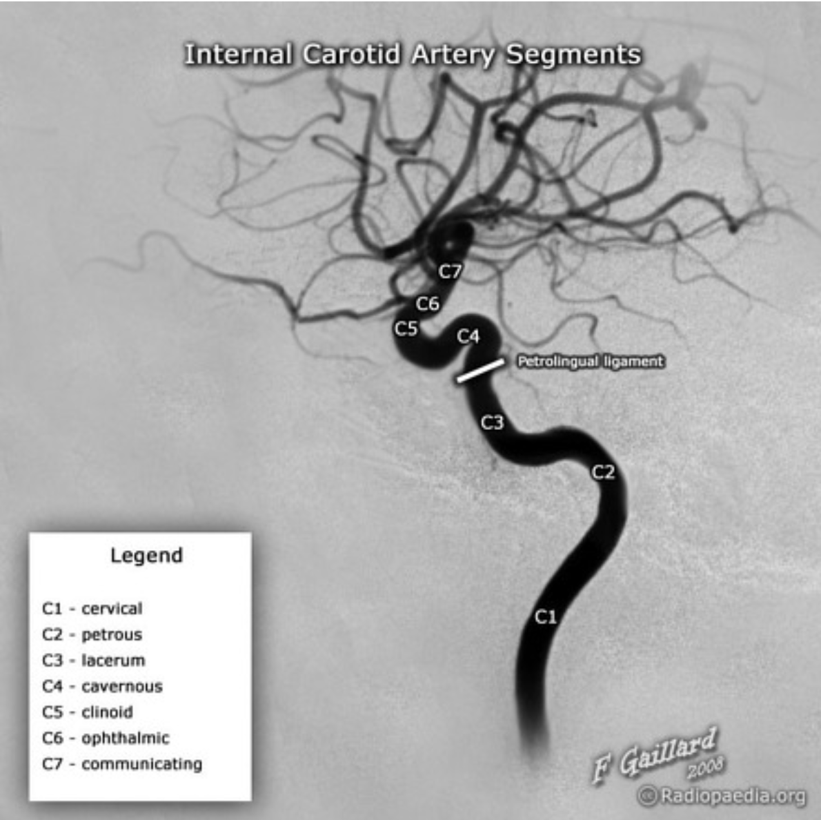
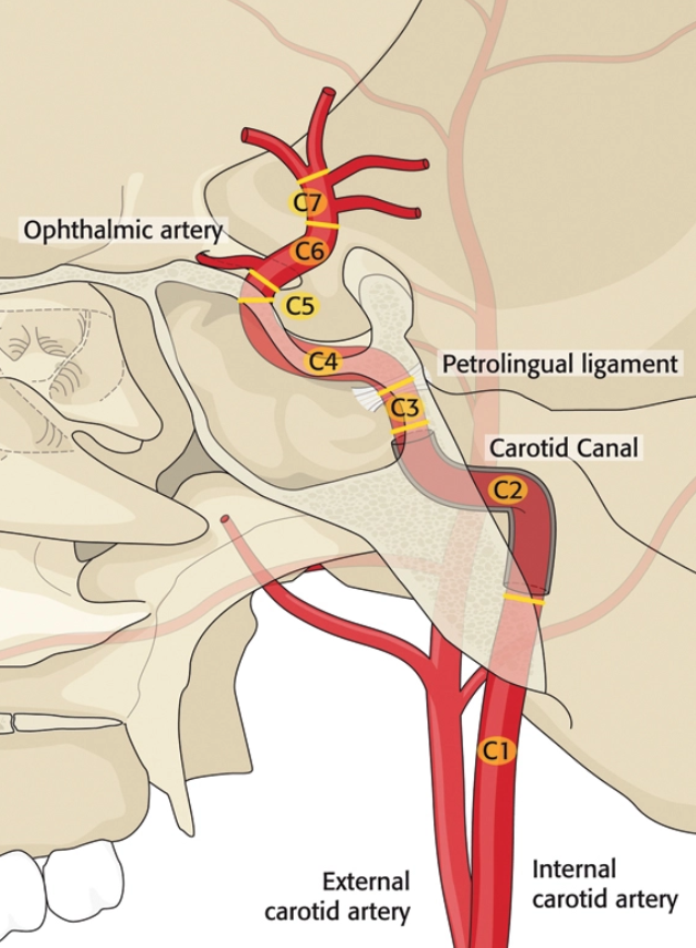
Internal Carotid Arteries
7 segments
Odd numbers = no branches (except 7)
Even numbers = have branches
C1 = cervical
C2 = petrous
Gives of vidian artery
C3 = lacerum
C4 = cavernous portion
Gives off Inferolateral trunk
Anastomoses with maxillary artery and can serve as collarteral flow from ECA
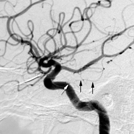
Gives off Meningohypophyseal trunk (black and white arrows in angio below)
Supplies hypophyseal trunk
C5 = clinoid
C6 = ophthalmic
Gives off ophthalmic artery
Gives off superior hypophyseal artery
C7 = communicating
External Carotid Artery
BIFL Grading
Grading system for vascular injury due to BLUNT trauma (does not apply to penetrating trauma such as GSW)
When discussing injury to the carotid arteries, Higher grade = higher risk of stroke
This same concept DOES NOT apply to other vessels
Grade 1:
Minimal luminal irregularity or
Intramural hematoma/dissection with <25% luminal narrowing
Grade 2: intramural hematoma, thrombus or dissection with ≥25% luminal narrowing
Grade 3: Pseudoaneurysm
Grade 4: Occlusion
Grade 5: Transection with extravasation
Artery of Adamkiewicz
Applies lower spine
Predominately on the left side and usually arises at about T7-L1
Ascends and makes hairpin turn where it anastomoses with the anterior spinal artery
Some correlation with embolization of bronchial artery embolization and bronchoesophageal fistula treatment because there may be connection between the fistula and spina arterial supply especially AoA so can accidentally embolize the Aoa
References:
Case courtesy of Disha Lokhandwala, Radiopaedia.org, rID: 161990 (PCA case)
Case courtesy of Disha Lokhandwala, Radiopaedia.org, rID: 163749 (MCA case)
Case courtesy of Disha Lokhandwala, Radiopaedia.org, rID: 162601 (ACA case)
Case courtesy of Jeremy Jones, Radiopaedia.org, rID: 32907 (Vert case)
Case courtesy of Frank Gaillard, Radiopaedia.org, rID: 36124 (Anterior circulation case)
Case courtesy of Frank Gaillard, Radiopaedia.org, rID: 36082