Neuro Trauma
Cerebral Contusions
Surface level insult to the cortical gray matter and some white matter deep to the affected area if enough force
Typically bilateral and multi-focal (90% of cases)
Will see new lesions, enlargement of lesions and hemorrhage typically within 24-48 hours
> 95% of cases will have extra-axial hemorrhage, most commonly SAH
Will have residual gliosis/encephalomalacia after healing
Most common location is anterior frontal lobes followed by temporal lobes
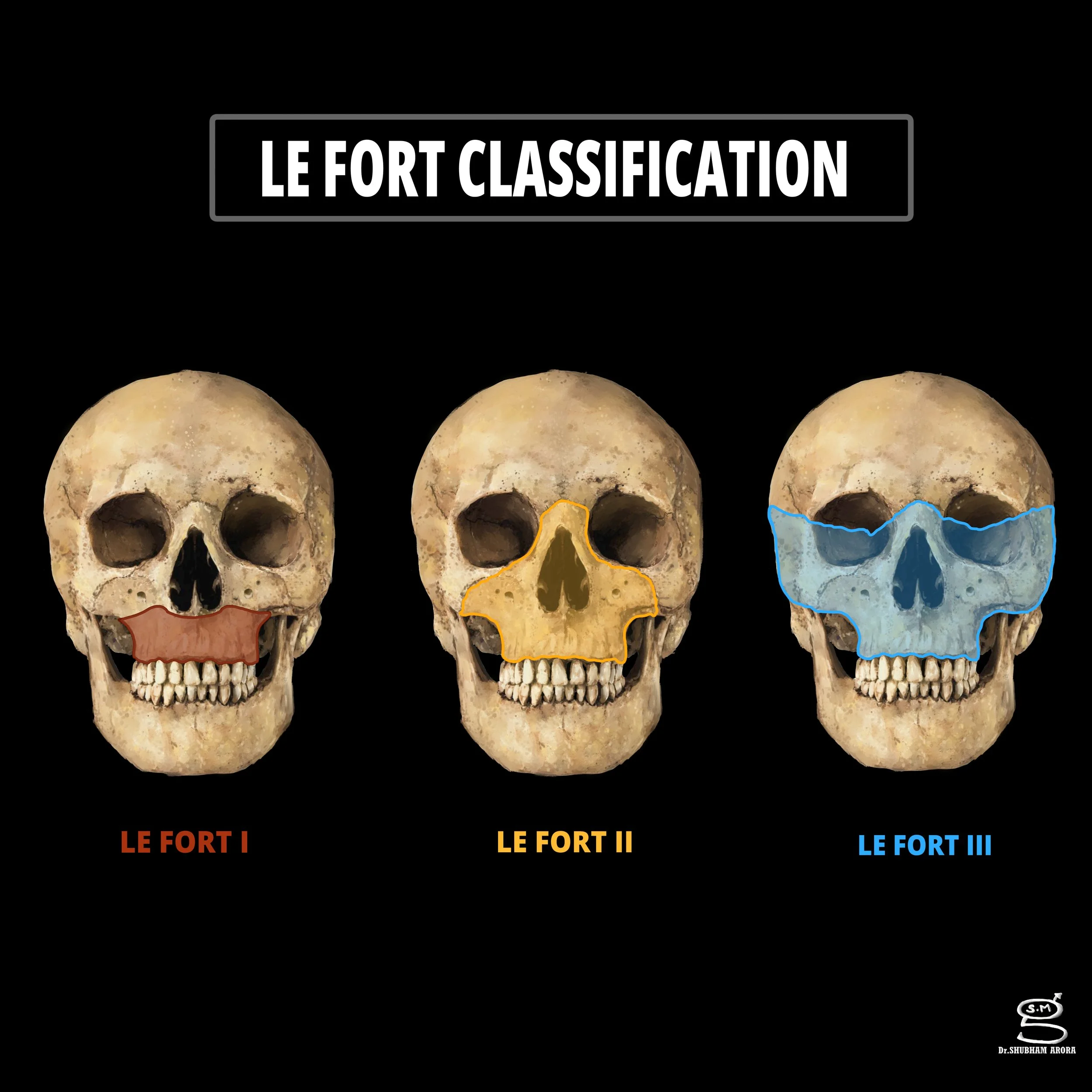
Le Fort Classification
Fractures of midface which result in separation of midface from skull base
Pteryoid plates and sphenoid bone always involved
Le Fort 1
Horizontal maxillary fracture separating teeth from rest of face
Key point - only one to involve anterolateral aspect of nasal fossa
Le Fort 2
Pyramid shaped fracture
Key point - only one to involve orbital rim
Le Fort 3
Key point - only one to involve zygomatic arch
Increased risk of temporalis muscle impingement
Highest risk of CSF leak
Kernohan Notch
Cerebral crus = white matter tracts at anterior aspect of cerebral peduncles
The Kernohan notch phenomenon occurs when there is a large midline shift which results in compression of the opposite cerebral peduncle (and therefore cerebral crus) which then results in a contralateral neuro deficit (because of the decussation), so
Expansile right sided lesion —> causes compression of left sided cerebral peduncle —> which causes a right sided deficit
Diffuse Axonal Injury
Caused by shearing forces
Multiple scattered hyperdense/hyperintense areas throughout brain in setting of trauma
3 Stages
Stage 1 - lesions at white matter/gray-white junction
Stage 2 - lesions in corpus callosum (+/- stage 1 findings)
Typically in splenium and body of CC
Stage 3 - lesions in brainstem (+/- stage 1 & stage 2 findings)
Hyperflexion spine injury
Widening of the posterior disc space
Widening of space between spinous processes
Focal kyphosis
Widened, jumped, perched facets
Whiplash
No imaging findings other than possible soft tissue edema
Hyperextension spine injury
Widening of the anterior disc space
Bony defect through posterior arch
Posterior element malalignment
Hyperextension + rotation spine injury
Focal vertebral body angulation
Rotation at site of injury
Resources: