MRI Basics
Terminology
Restriction (Restricted diffusion)
Means that there is a decreased amount of flow (diffusion) of water molecules into a tissue
Lets do a thought experiment…
Imagine a large hallway measuring 30 feet or so. In this hallway there are 30 people lined up all walking through the hallway at the same time without any collisions or difficulty. Now this would be normal diffusion. So what would make us have a decrease in the number of people who can walk through the same hallway? Well there’s 2 maybe 3 reasons why this could happen.
Reason 1:You decrease the size of the hallway. If all 30 people passed through the hallway perfectly to begin with and we make the hallway 2X smaller then half the people would be able to pass at a time. This means less people than normal can pass through the hallway at any given time, which in radiology terms means less flow of water to a tissue = restricted diffusion.
Reason 2: You increase the size of the people trying to get through the hallway. If the 30 people fit perfectly through the hallway originally then if you make each individual 2X bigger then they would not all be able to get through the hallway at once and therefore some people would have to wait until the first round of people went through before they could pass. This means less people are going through the hallway at any given point in time, or in other words there is less flow of water into a tissue = restricted diffusion. This is the equivalent of increasing particle size which is not really as relevant as other example for radiology purposes.
Reason 3: We kill all the people and there is just a space and no people to move through the hallway. - This is somewhat of an exception but demonstrates what we seen in a chronic stroke. If there is no T2 signal at all then you cannot have flow of water but because there is essentially nothing there then it doesn’t count and does not follow the same rules or appearance of restricted diffusion and will just appear black.
Hyperintense on DWI (diffusion weighted imaging)
Indicates low ADC (apparent diffusion coefficient)
Restricted diffusion typically indicates that there is hypercellularity
General principle to help remember reasons why certain disease cause restricted diffusion
If a cell is dying or does not have enough energy to function then the machinery and transporters in the cell wall will stop working and cannot take in water which results in decreased flow of water = restricted diffusion
Infarct = cell is dying and the transporters are no longer functioning
Hypoglycemia = no energy to run the transporters
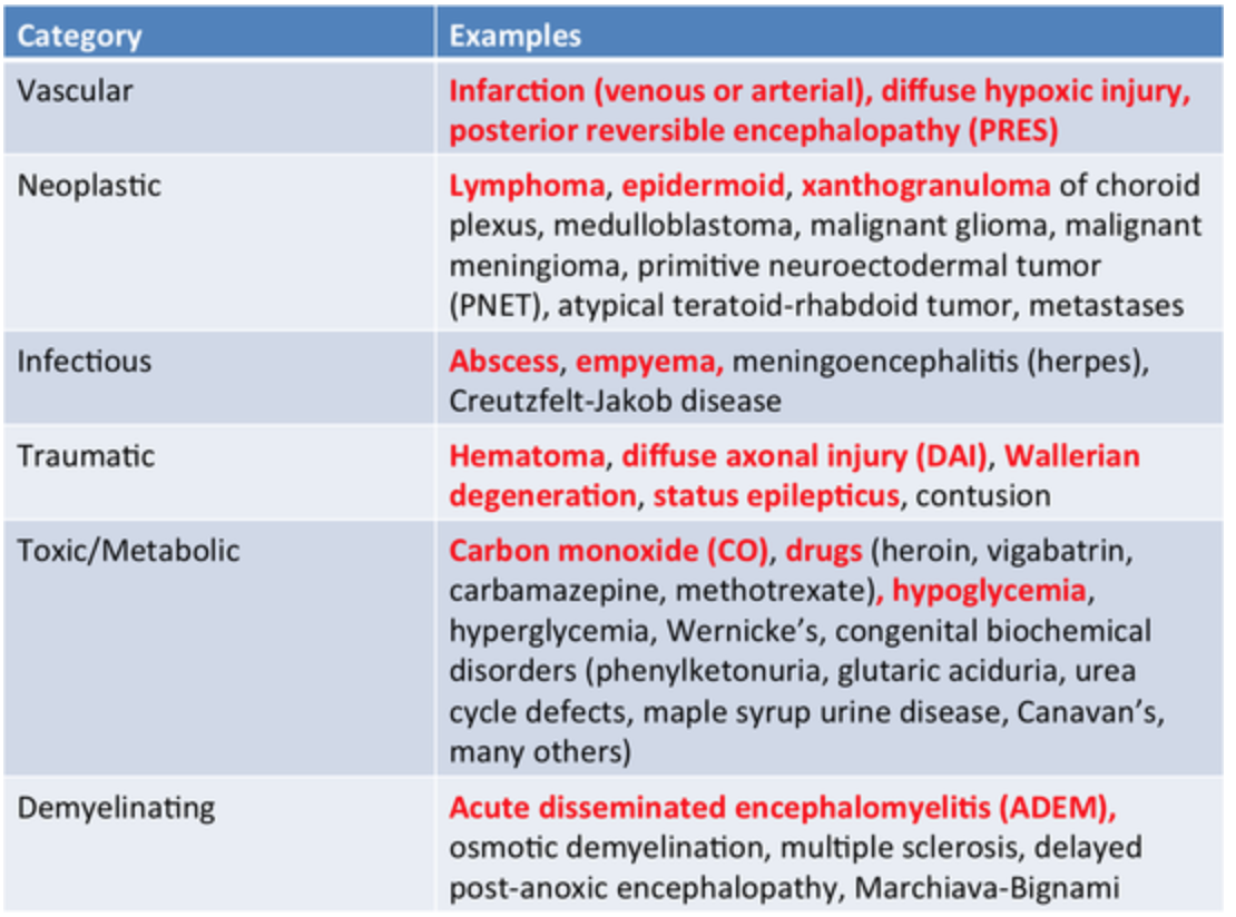
Most important causes of Restricted Diffusion
Infarction
Abscess
Lymphoma
Myelin edema
T1 Weighted Imaging
Utilizes difference in longitudinal relaxation times (1 in T1 looks like L for longitudinal)
Short (echo time) TE of ~ 10-20 ms
Short (repetition time) TR of ~ 400-600 ms
Fat is bright/hyperintense
Water is dark/hypointense
T2 Weighted Imaging
Utilizes difference in transverse relaxation times)
Long echo time, TE ~ 90-110 ms
Long repetition time, TR ~ 3000-6000 ms
Fluid is bright/hyperintense
Proton Density
Utilizes density of hydrogen protons in the tissue
Long TR, 2000 ms < (allows for complete relaxation of longitudinal magnetization before next pulse)
Short TE, <20 ms (allows you to capture signal before transverse relaxation occurs)
Fat - bright/hyperintense
FLAIR
Utilizes inversion time
Inversion time typically chosen in 2000-2500 range ms to adequately suppress CSF
Long TR, 6000 ms <
Mid TE, ~100 ms
Suppresses CSF signal
Fat - bright/hyperintense
Fluid (CSF) - dark/hypointense
References: