Neuro Metabolic Disease
Cerebral Amyloid Angiopathy
Blooming black dots
Seen in amyloid disease and chronic hypertensive encephalopathy
May resemble chronic hypertensive encephalopathy on imaging
Both will have blooming black dots
In cortex and pia —> amyloid
In basal ganglia and cerebellum —> HTN
Superficial siderosis present
Systemic siderosis does not correlate with CAA
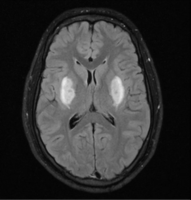
Eye of the Tiger Sign
Symmetric low T2 signal of globus pallidus (due to accumulation of iron) with central area of high signal (due to gliosis)
Associated with PKAN (pantothenate kinase associated neurodegeneration)
Other associations
Organophosphate poisoning
Parkinsonism
Wilson disease
Eye of the Tiger Sign
Symmetric low T2 signal of globus pallidus (due to accumulation of iron) with central area of high signal (due to gliosis)
Associated with PKAN (pantothenate kinase associated neurodegeneration)
Other associations
Organophosphate poisoning
Parkinsonism
Wilson disease
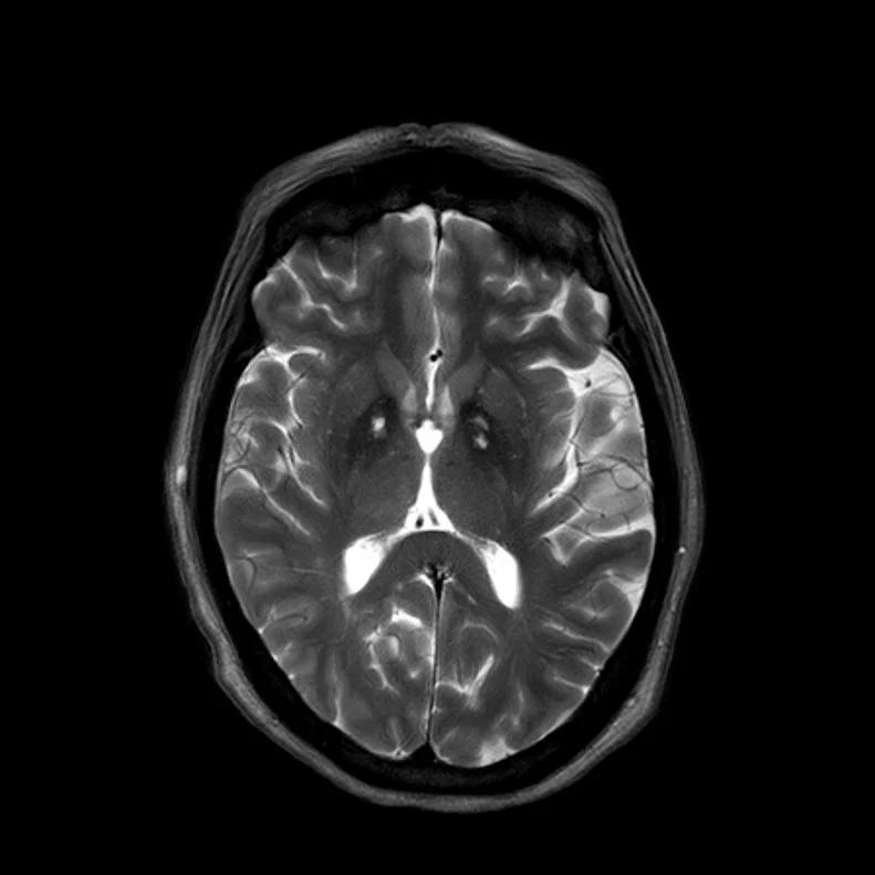
Mitochondrial encephalomyopathy with lactic acidosis and stroke-like episodes (MELAS)
Multifocal stroke like lesions in different stages of healing and does not follow a specific vascular territory
Prefers posterior parietal and occipital lobes
Seen < 40 yo
Black toenail sign
Basal Ganglia
Bilateral Basal Ganglia
T1 Bright basal ganglia
Liver failure
Hyperalimentation (manganese)
High blood sugar (non DKA)
Wilsons disease
Viral infection (West Nile, Japanese Encephalitis)
Drugs
Heroin
CO poisoning
Methanol
Cyanide
Niroimidazole
Methanol Poisoning
Classically Symmetric Putamen hyperintensity
Looks like orange slice candy
Nonspecific bilateral - typically symmetric basal ganglia involvement
Cyanide
Organophosphate poisoning
Typically lacks hemorrhage which may be seen with others
Carbon Monoxide Poisoning
Classically Symmetric globus pallidus hyperintensity
Thalamus
Bilateral Thalami Involvement
Wernickie
Artery of Percheron infarct
Internal cerebral vein thrombosis
Wernickie Encephalopathy
Caused by thiamine deficiency typically in alcoholics
Findings
T2/FLAIR hyperintensity of the bilateral thalami and periaqueductal gray matter
Mammillary body enhancement
References:
Case courtesy of Yune Kwong, Radiopaedia.org, rID: 41008
Case courtesy of Elshan Abdullayev, Radiopaedia.org, rID: 50288