Liver Ultrasound
Basics
Types of Ultrasound
Gray scale
Color doppler
Spectral Doppler
Doppler angle
Angle formed between doppler waves and the structure of interest
Need to be <60 deg (bunch of physics shit for this)
Terminology
Terms referring to anatomy
Antegrade flow
Blood flowing forward in normal circulatory flow pattern
i.e - IVC = toward the heart, aorta, away from heart, etc.
Retrograde flow
Blood flowing backward in opposite direction of normal flow pattern
Terms referring to probe/transducer location
Toward transducer —> red —> above line
Away from transducer —> blue —> below line
Key point
Ante-grade flow does not mean toward the transducer, you can have antegrade flow that is away from transducer and vice verse, same goes for retrograde flow
Phasicity
Basically how many different shapes there are within a wave
You can get lost in the terms here which I doubt really matter much, beyond maybe aphasic which is just a flat line
Spectral broadening
Area under the curve - how sharp a line is - something like this idk should relook at this
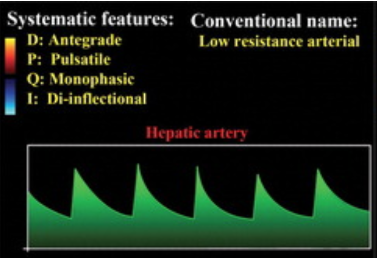
Hepatic Artery
Pulsatile
Antegrade throughout entire cycle —> therefore entire waveform should be above the line
Peak = peak systolic velocity
Trough = end diastolic velocity
Low RI = 0.55-0.7
Reasons for increased RI —> postprandial, old age, peripheral microvascular disease, hepatic venous congestion, etc.
Reasons for decreased RI —> HHT, arcuate ligament syndrome, cirrhosis with portal HTN,
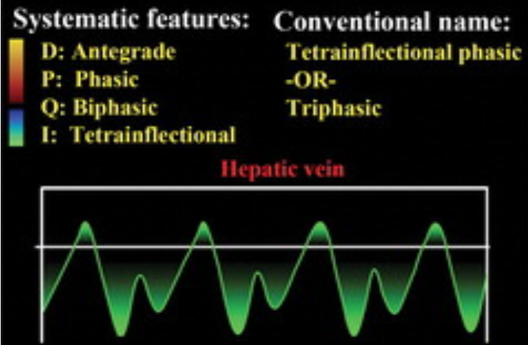
Hepatic Veins
H
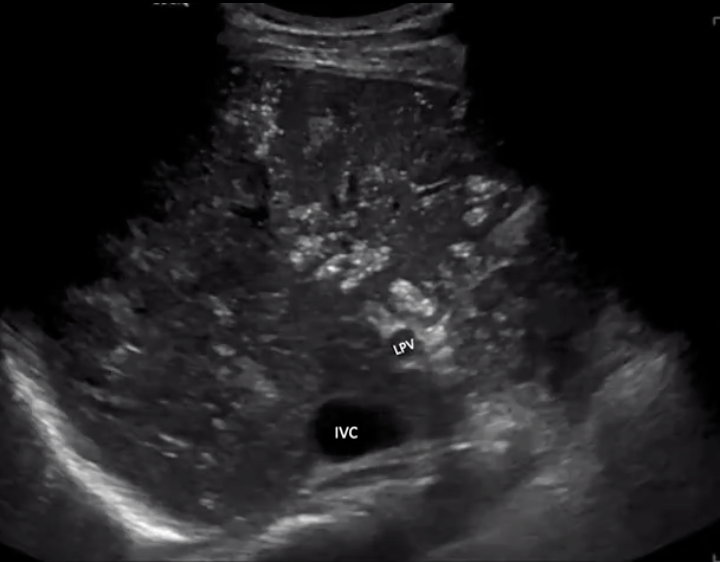
Portal Vein
Normal Characteristics
Hepatopetal flow (toward the liver)
Should make sense because portal system is basically its own closed circuit of veins and exchanges at the liver so should not be hepatofugal because, well the portal vein does not go to the heart
Antegrade (above the line)
Can have gentile undulations (wavy appearance)
Normal velocity = 16-40
Normal size = <13 mm
Steatosis
Increased liver echogenicity (brighter)
Multiple stages - no body cares except body imagers
Cirrhosis
Nodular contour
Coarse and heterogenous echotexture
Enlarged portal vein with decreased portal vein velocity
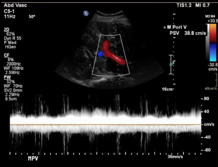
Portal Venous Gas
Echogenic foci which extend to the periphery of the liver
Key way to differentiate from pneumobilia which is more centrally located
Commonly see gas near portal vein
Bidirectional spikes on doppler (see below)
DDx
Bowel perforation
Bowel ischemia
Recent surgery
Others…
References: