Eye
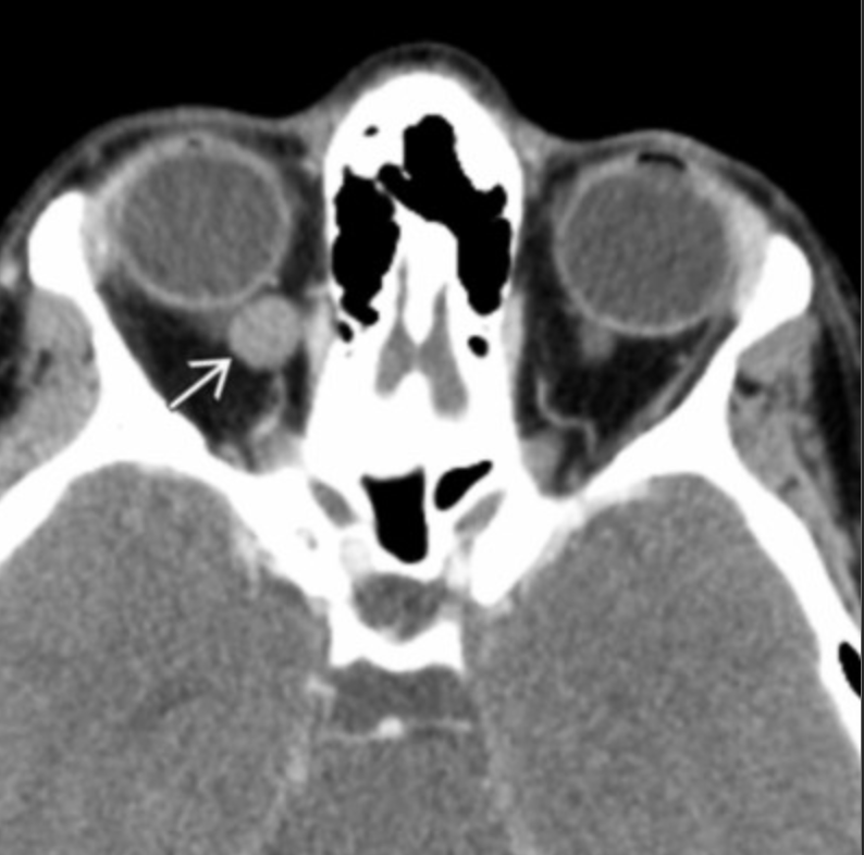
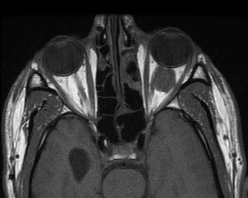
Orbital Cavernous malformation
Basically hemangioma of the eye - but no true epithelial capsule and technically not a neoplasm
>80% are intraconal, typically laterally located
Presents with proptosis and can cause local compression - eye issues
Surgical resection is curative
Rarely hemorrhage (note venolymphatic malformations in kids do have hemorrhage)
Coats Disease (Retinal telangiectasias)
Typically age 3-9
M>F
Affected eye is smaller than normal eye
May see retinal detachment (V-shape)
Unilateral
Basically looks like a single messed up eye
Retinoblastoma
95% of cases diagnosed before age 5 yo
Presents with leukocoria
Calcified retinal lesions with increased vascularity on doppler
Unilateral or bilateral (auto dominant inheritance typically for bilateral disease)
Trilateral Rb
Bilateral Rb (each eye) + Pineoblastoma
Quadrilateral Rb
Trilateral + suprasellar embryonal CNS tumor + osteosarcoma
Mets
via Optic nerve cuasing leptomeningeal enhancement
also to bone
Tolosa Hunt Syndrome
Angiolymphatic malformation
Multiloculated cystic and solid mass
Possible adjacent osseous remodeling secondary to persistent compression on bone
Really just looks like some fuck shit behind the eye
Fluid-fluid levels (Rb does not have)
Less commonly have calcs (Rb has calcs)
Extra and intraconal
Proptosis and swelling (Rb may have some of this but leukocoria and vision loss is key)
Resources: