Esophagus
Normal Esophageal Anatomy
Normal esophageal width = 3 mm
Upper 1/3 = striated muscle
Middle 1/3 = mixed striated and smooth muscle
Lower 1/3 = smooth muscle
Extends from cricopharyngeus muscles to the stomach
Malignant Esophageal Masses
Malignant masses can easily invade local structures due to lack of serosal covering of the esophagus
Look for —> Esophageal wall thickening + Intraluminal mass + Lumen narrowing
Squamous cell carcinoma (75%)
Adenocarcinoma (25%)
Secondary to Barrett’s esophagus and malignant transformation of dysplastic tissue
Benign Esophageal Masses
Well circumscribed mass
These are the only esophageal masses that can have calcifications (more so leiomyoma than GIST)
Leiomyoma
Tend to be large (>5 cm)
Not FDG-avid
GIST
FDG-avid
Papilloma
Most common benign lesion
Esophageal Diverticula
Feline Esophagus
Associated with reflux esophagitis
Folds are only in lower 2/3 of esophagus
Folds are transient and go away with swallowing
Esophageal Pseudodiverticulosis
Basically dilated excretory mucosal ducts
Look lie a bunch of squiggly lines along the borders of the esophagus
Associated with
esophageal structures - 90%
Reflux
Candidiasis
Candida esophagus
Seen in
Immunocompromised (GHIV, transplant)
Motility disorders (scleroderma, achalasia)
Presents as plaque like lesions, may be shaggy if severe
Herpes
Small and multiple ulcers with halo of edema
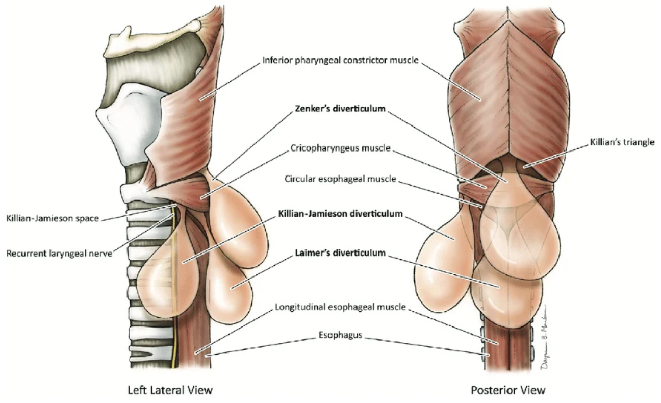
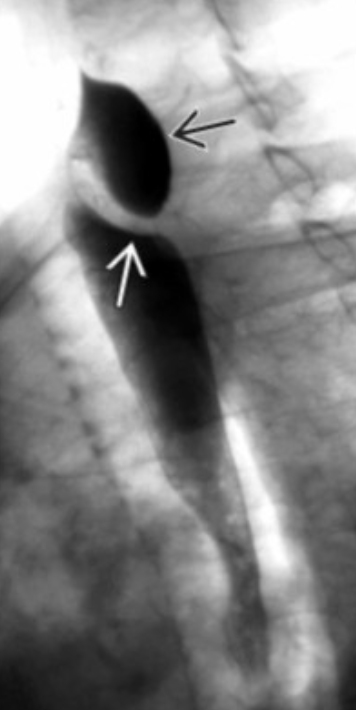
Zenker Diverticulum
Posterior
Just above cricopharyngeus muscle
In the hypopharynx
Area of weakness is the Killian triangle (but not a killin diverticula, nonsense I know)
Killian-Jamieson Diverticula
Anterolateral wall of cervical esophagus
Below cricopharyngeus muscle
Usually smaller and obv anterior compared to zenker
Rarest form of esophageal diverticula
Classic Esophagram Lesions
Esophageal Non-neoplastic lesions
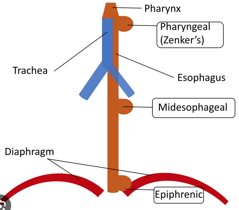
Traction Diverticula
Seen at mid-esophagus
Said to be triangular, looks round to me
Occur secondary to scarring (prior granulomatous disease)
Epiphrenic Diverticula
Basically just above the GE junction at the diaphragm, hence “phrenic”
Typically on the RIGHT
Note, para-esophageal is typically on left
Look
Esophageal Web
Most common in cervical esophagus
Increased risk of esophageal and hypopharyngeal cancer
Plummer-Vinson
Iron deficiency anemia
Dysphagia/Esophageal web
Thyroid issues
Spoon shaped nails
Glycogenic Acanthosis
Basically focal collections of glycogen
Presents basically the same as Candida but
Seen in old people who are otherwise normal (not immunocompromised)
Asymptomatic
Esophageal Ulcers
CMV & HIV
Large and flat ovoid ulcers
Crohns
Rare unless severe disease
Look for description as Aphthous with prominent surrounding edema
References: