High Yield Signs for Chest Radiography
Atoll Sign
Bulging fissure sign
Continuous diaphragm sign
Crazy paving
Sabre sheath sign
(Retrocardiac) Sail sign
Hour glass sign
Horseshoe lung
Hilum overlay sign
Hoffman-Rigler Sign
Deep sulcus sign
Double density sign
Running man sign
Finger in glove
Golden “S” sign
H convergence sign ?
Luftsichel sign
Split pleura sign
Snoopy Sign
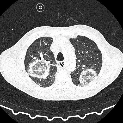
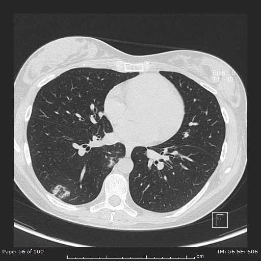
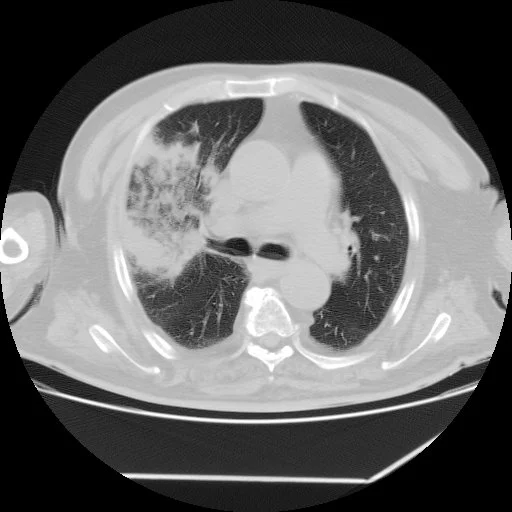
Atoll Sign (Reverse Halo Sign)
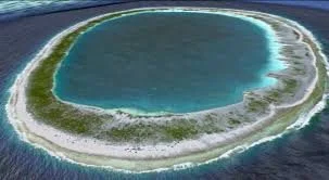
Atoll = circular island with water in the middle (looks like a doughnut)
Central ground glass opacity with consolidative/enhancing rim
DDx:
Cryptogenic organizing pneumonia - main cause
Fungal pneumonia
Wegeners (granulomatosis with polyangitis)
Sarcoid or TB
Others
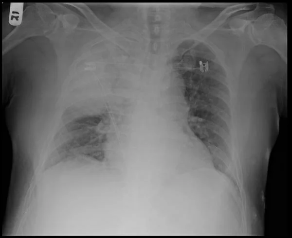
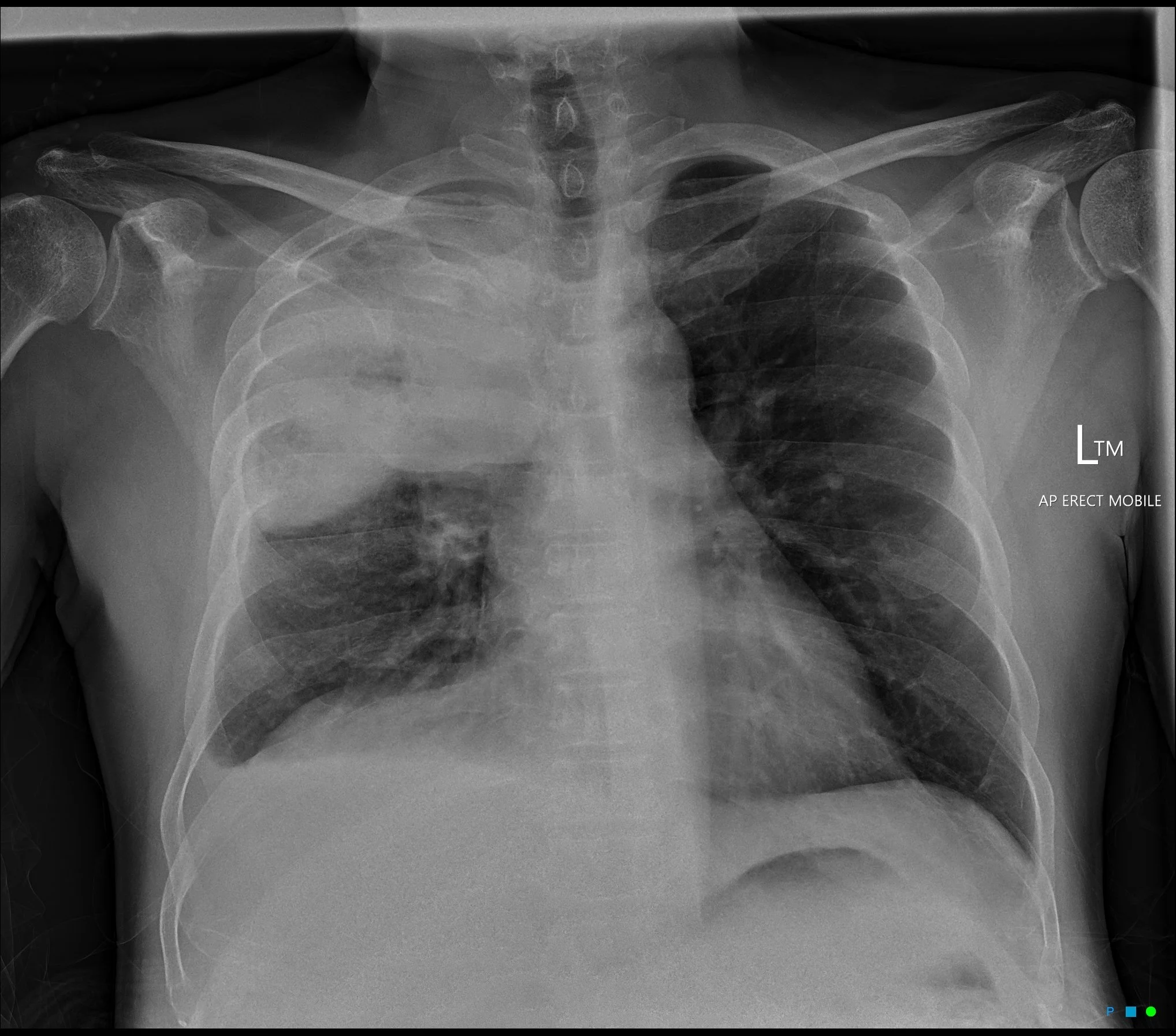
Bulging Fissure Sign
Consolidation with expansion of the lung lobe such that the fissure boundary is displaced
Caused by anything that occupies space and therefore exerts mass effect within the lung
DDx:
Pneumonia
Classically seen in RUL secondary to Klebsiella pneumonia
Strep pneumoniae, Staph aureus, Pseudomonas too
TB
Legionella
Cancer (Adenocarcinoma of the lung)
Pulmonary hemorrhage
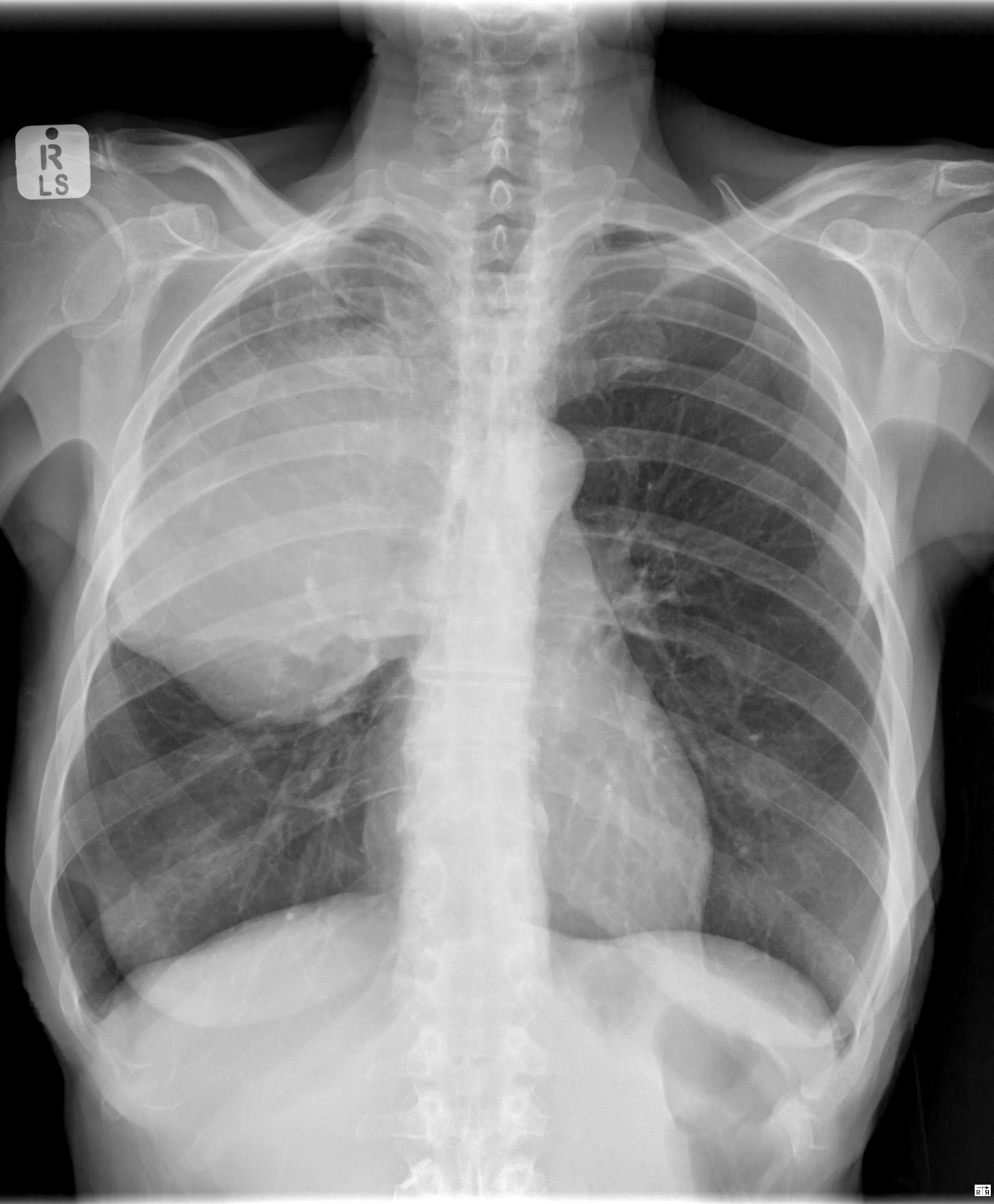
Sabre Sheath Sign
Narrowing of intrathoracic portion of trachea (basically looks like trachea gradually tapers) on CXR
On cross sectional imaging will look longer (in AP) than wider
Pathognomonic for COPD
Not sure I can explain how exactly this looks like a sabre-sheath but…
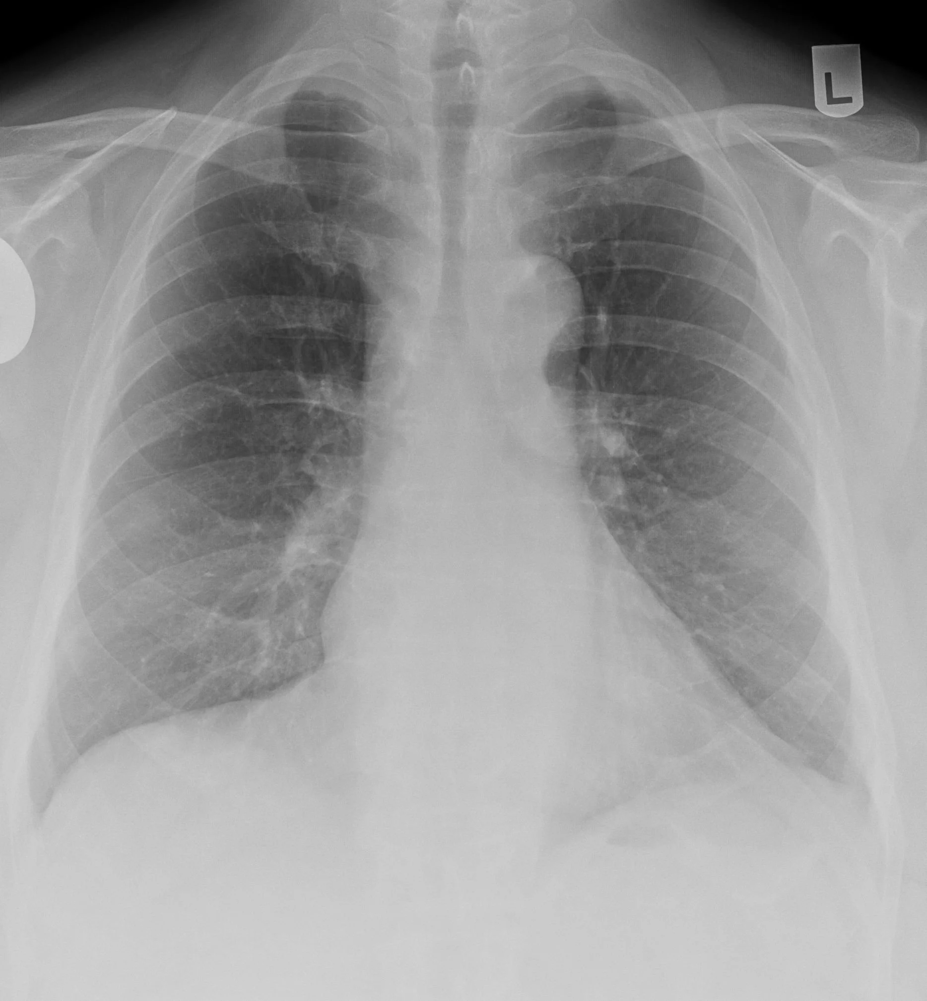
Snoopy Sign
Seen in pericardial agenesis
Will see
Elongated and protruding left heart border
Subtle lucency between the pulmonary artery and aorta (because no longer have pericardium which extends superiorly to bridge the two so you are basically seeing the borders of the vessels clearly
Lucency between left heart border and diaphragm
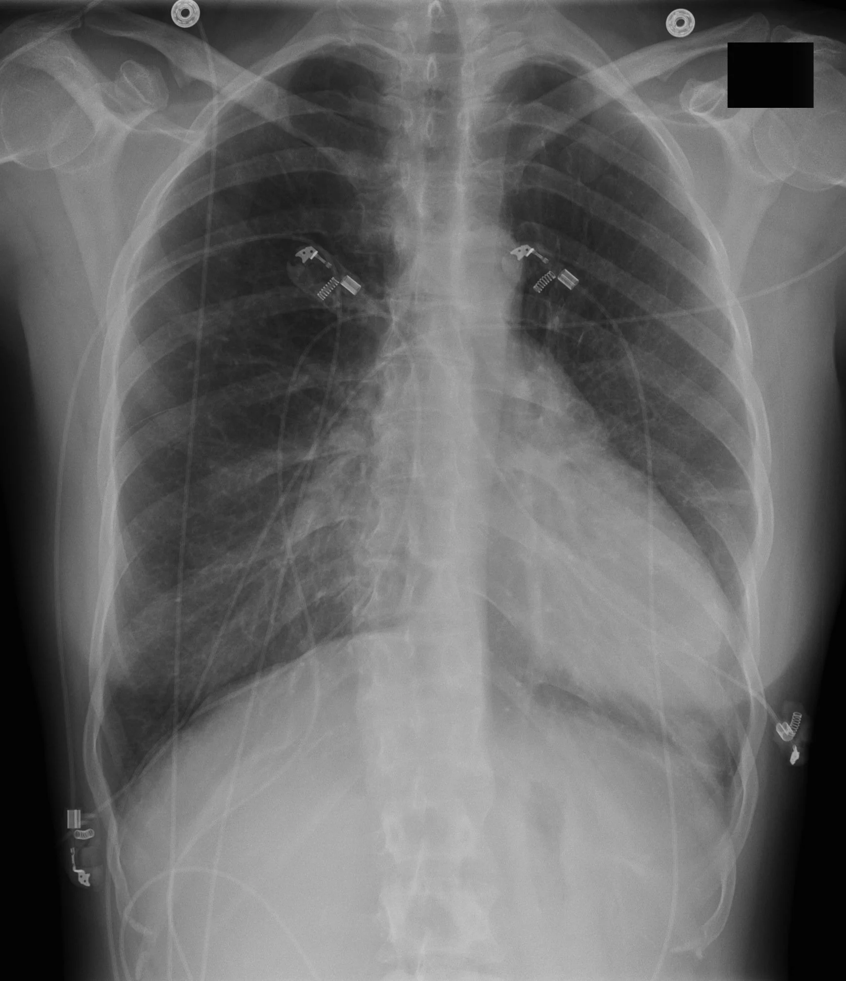

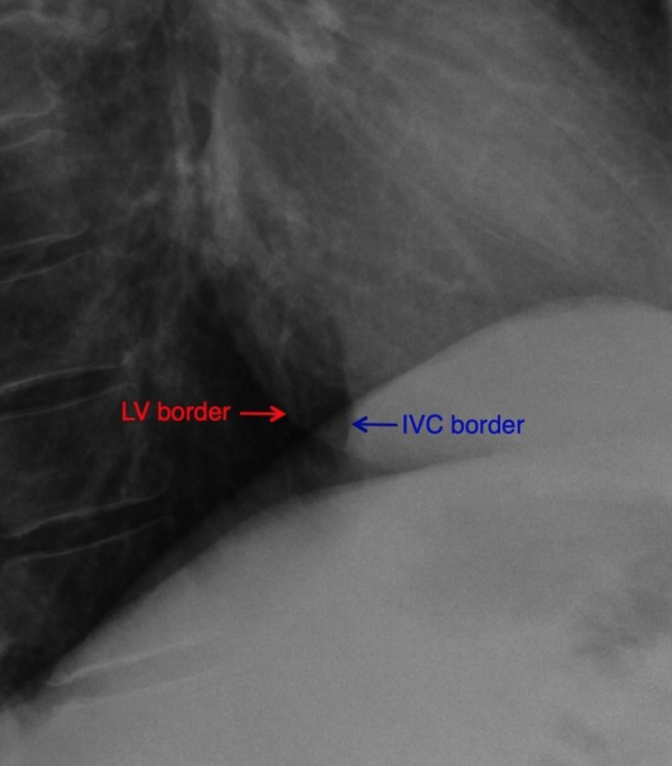
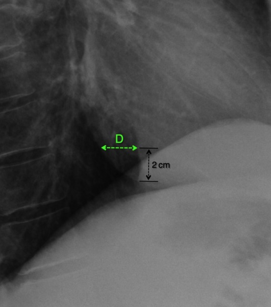
Hoffman-Rigler Sign
Seen in LV hypertrophy
Only seen on lateral radiograph and measured 2 cm above the intersection of IVC and diaphragm
Distance between the LV border and posterior border of IVC is >2.8cm
References:
Case courtesy of Raad Al Tahat, Radiopaedia.org, rID: 82033 (Atoll sign)
Case courtesy of IRSHAD AHMAD PAUL, Radiopaedia.org, rID: 183961 (Atoll sign)
Case courtesy of Eid Kakish, Radiopaedia.org, rID: 87856 (Atoll sign)
Case courtesy of Ashesh Ishwarlal Ranchod, Radiopaedia.org, rID: 176761 (bulging fissure)
Case courtesy of Craig Hacking, Radiopaedia.org, rID: 79675 (bulging fissure)
Case courtesy of The Radswiki, Radiopaedia.org, rID: 11266 (bulging fissure)
Case courtesy of Andrew Ho, Radiopaedia.org, rID: 28522 (sabre sheath)
Case courtesy of Maxime St-Amant, Radiopaedia.org, rID: 20695 (snoopy sign)
Case courtesy of Vincent Tatco, Radiopaedia.org, rID: 44892 (Hoffman Rigler)