Bowel Disease
General
Richter Hernia (Parietal Hernia)
Basically hernia but only part of the bowel wall is herniated and not the whole like (like a pseudo-hernia almost because all layers of bowel are not involved)
Most commonly seen in femoral ring
Higher chance of gangrene and less chance of obstruction
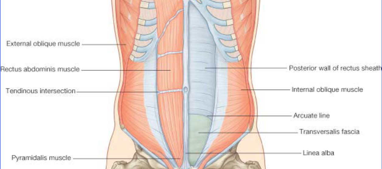
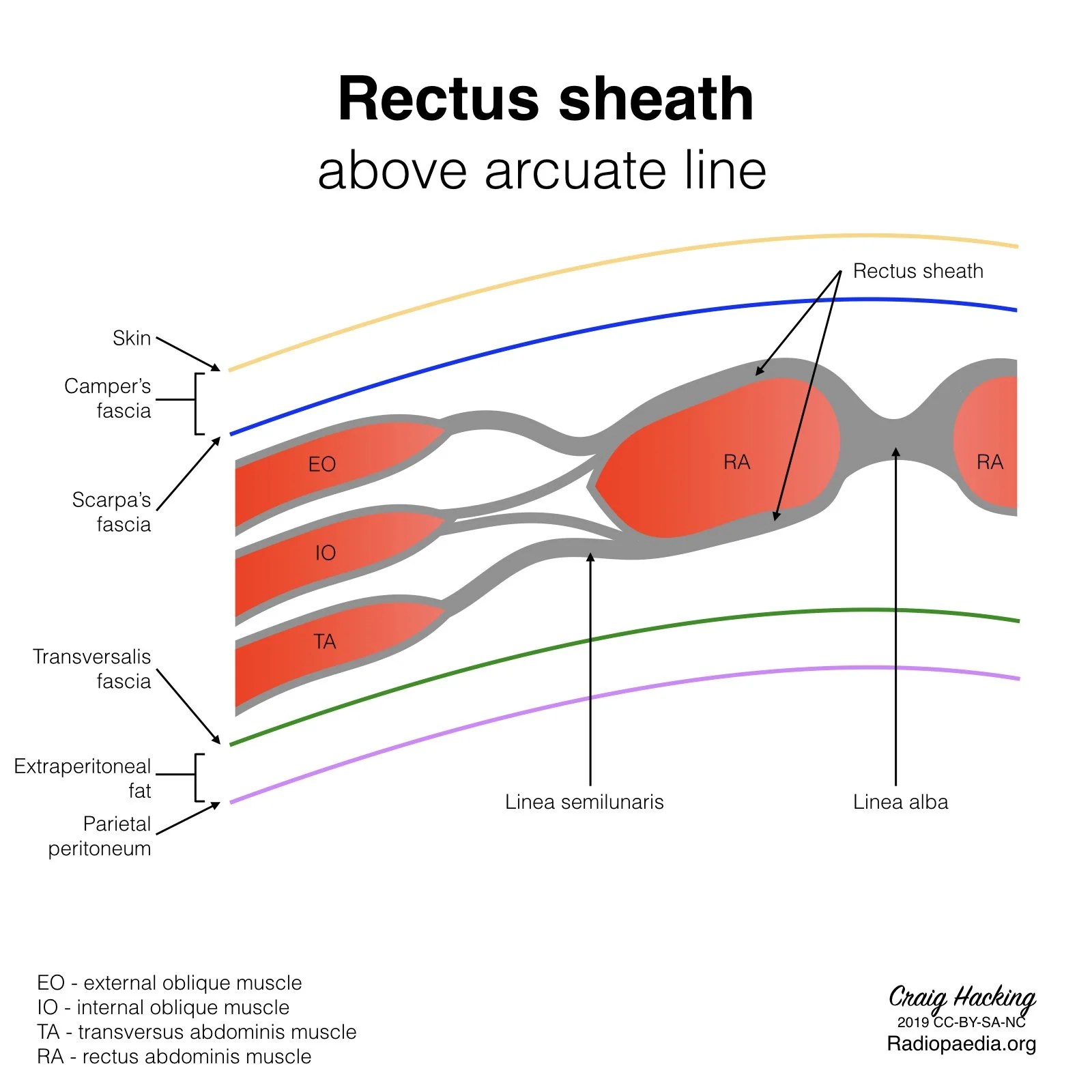
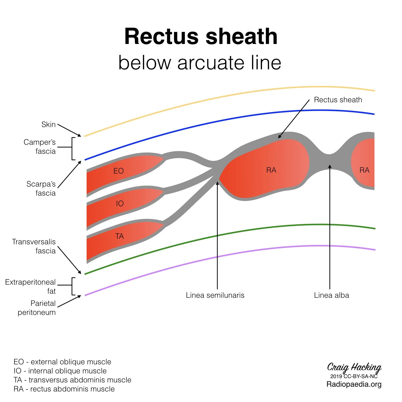
Spigelian Hernia
Hernia through defect of internal oblique and transverse abdominus muscle aponeurosis
Location
Inferior and lateral to umbilicus
Will be at or below level of arcuate line = no posterior rectus sheath at this level, TA and IO fibers run parallel here
Deep to external oblique aponeurosis
Metabolic Disease
Evaluation
Supine - air may not be seen in colon due to positioning
Prone - air should be visible in colon
Crohns
Wall thickening and enhancement
3-5 mm
5-9 mm
10+ mm
Comb sign - thickened vasa recta extend from thick bowel wall looking like a comb
Skip lesions - normal bowel interspersed with diseased bowel
Fistulas and Abscesses form
Celiac Disease
Dilated, thickwalled and fluid filled loops of small bowel
Jejunization of the ileum (increased bowel wall folds in the ileum and decreased wall folds in the jejunum)
Small Bowel Obstruction
Complete SBO
Dilated proximal small bowel and collapsed distal bowel and colon with transition point
Transition point = where bowel goes from dilated to collapsed
Ileus
Dilated proximal and distal small bowel with no transition point
Partial small bowel obstruction
Somewhere in between complete SBO and ileus
Bowel is not as distended and transition point is not as definitive
Fecalization of the small bowel
Delayed transit in small bowel results in increasd water resorption and appearance of fecal material in the small bowel lumen
Seen in states of delayed transition, including partial SBO
Closed loop obstruction
Piece of bowel occluded at proximal and distal ends - think of a sausage link
Can twist resulting in volvulus
Sclerosing Mesenteritis
Affects jejunal mesentery
Clusters of mesenteric nodules
Desmoid Tumor
Locally aggressive mass
Associated with Gardner syndrome
Throughout abdomen and abdominal wall
Look like homogenous soft tissue masses in abdomen
T1 & T2 hypointense
Variable enhancement
Enteric Duplication Cyst
Water density cyst arising from GI tract
Most common location
#1 = Ileum
#2 = Esophagus (posterior mediastinal cyst)
Masses
Intestinal (Angioneurotic) Angioedema
Basically angioedema like you get in the face but in the bowel
Bowel wall edema, ascites
Associated with ACEi use
Hernias
Carcinoid Tumor
Desmoid Tumor
Resources:
Case courtesy of Craig Hacking, Radiopaedia.org, rID: 67538 (arcuate line)